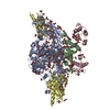
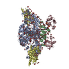
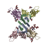
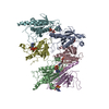
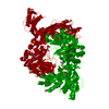
- PDB-5dvy: 2.95 Angstrom Crystal Structure of the Dimeric Form of Penicillin... -
+
Open data
ID or keywords:
Loading...
-
Basic information
Entry
Database: PDB / ID: 5dvy
Title
2.95 Angstrom Crystal Structure of the Dimeric Form of Penicillin Binding Protein 2 Prime from Enterococcus faecium
Components
Penicillin binding protein 2 prime
Keywords
PENICILLIN-BINDING PROTEIN / penicillin binding protein 2 prime / PBP2 / CSGID / Structural Genomics / Center for Structural Genomics of Infectious Diseases
Type: MARMOSAIC 225 mm CCD / Detector: CCD / Date: Mar 9, 2015
Radiation
Monochromator: C(111) / Protocol: SINGLE WAVELENGTH / Monochromatic (M) / Laue (L): M / Scattering type: x-ray
Radiation wavelength
Wavelength: 0.97872 Å / Relative weight: 1
Reflection
Resolution: 2.95→30 Å / Num. obs: 36548 / % possible obs: 99.8 % / Observed criterion σ(I): -3 / Redundancy: 13.2 % / Biso Wilson estimate: 68.2 Å2 / Rmerge(I) obs: 0.13 / Rsym value: 0.13 / Net I/σ(I): 23.7
Reflection shell
Resolution: 2.95→3 Å / Redundancy: 13.5 % / Rmerge(I) obs: 0.69 / Mean I/σ(I) obs: 4 / % possible all: 100
-
Processing
Software
Name
Version
Classification
REFMAC
5.8.0073
refinement
HKL-3000
datareduction
HKL-3000
datascaling
PHENIX
phasing
BLU-MAX
datacollection
Refinement
Method to determine structure: SAD / Resolution: 2.95→29.91 Å / Cor.coef. Fo:Fc: 0.961 / Cor.coef. Fo:Fc free: 0.942 / SU B: 14.762 / SU ML: 0.144 / Cross valid method: THROUGHOUT / ESU R: 0.284 / ESU R Free: 0.218 / Stereochemistry target values: MAXIMUM LIKELIHOOD / Details: HYDROGENS HAVE BEEN ADDED IN THE RIDING POSITIONS
Rfactor
Num. reflection
% reflection
Selection details
Rfree
0.18678
1762
4.8 %
RANDOM
Rwork
0.1527
-
-
-
obs
0.15437
34772
99.45 %
-
Solvent computation
Ion probe radii: 0.8 Å / Shrinkage radii: 0.8 Å / VDW probe radii: 1.2 Å / Solvent model: MASK
 Movie
Movie Controller

 Yorodumi
 Open data
 Basic information
 Components
 Keywords
 Function and homology information
 Enterococcus faecium DO (bacteria)
 X-RAY DIFFRACTION /
  Authors
 Citation
 Structure visualization
 Downloads & links
 Other downloads
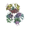

 PDBj
PDBj

 Assembly
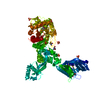

 Mass: 122.143 Da / Num. of mol.: 1 / Source method: obtained synthetically / Formula: C4H12NO3 / Comment: pH buffer*YM
 Mass: 96.063 Da / Num. of mol.: 14 / Source method: obtained synthetically / Formula: SO4
 Mass: 18.015 Da / Num. of mol.: 243 / Source method: isolated from a natural source / Formula: H2O
 Sample preparation
 / Beamline: 21-ID-F / Wavelength: 0.97872 Å
 Processing