 Movie
Movie Controller

[English] 日本語
 Yorodumi
- PDB-6i2p: Crystal structure of the Mycobacterium tuberculosis PknB kinase d... -
+ Open data

- Basic information
| Entry | Database: PDB / ID: 6i2p | ||||||
|---|---|---|---|---|---|---|---|
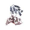
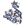
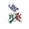
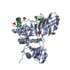
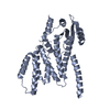
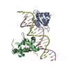
| Title | Crystal structure of the Mycobacterium tuberculosis PknB kinase domain (L33E mutant) in complex with its substrate GarA | ||||||
 Components Components |
| ||||||
 Keywords Keywords | SIGNALING PROTEIN / kinase / FHA-domain protein | ||||||
| Function / homology |  Function and homology information Function and homology informationnegative regulation of growth rate / negative regulation of fatty acid biosynthetic process / acetyltransferase activator activity / regulation of cellular respiration / negative regulation of glycolytic process / molecular function inhibitor activity / peptidoglycan biosynthetic process / peptidoglycan-based cell wall / regulation of cell shape / manganese ion binding ...negative regulation of growth rate / negative regulation of fatty acid biosynthetic process / acetyltransferase activator activity / regulation of cellular respiration / negative regulation of glycolytic process / molecular function inhibitor activity / peptidoglycan biosynthetic process / peptidoglycan-based cell wall / regulation of cell shape / manganese ion binding / protein kinase activity / non-specific serine/threonine protein kinase / protein serine kinase activity / protein serine/threonine kinase activity / mRNA binding / regulation of DNA-templated transcription / extracellular region / ATP binding / identical protein binding / plasma membrane Similarity search - Function | ||||||
| Biological species |   Mycobacterium tuberculosis (bacteria) Mycobacterium tuberculosis (bacteria) | ||||||
| Method |  X-RAY DIFFRACTION / SYNCHROTRON / MOLECULAR REPLACEMENT / Resolution: 2.37 Å X-RAY DIFFRACTION / SYNCHROTRON / MOLECULAR REPLACEMENT / Resolution: 2.37 Å | ||||||
 Authors Authors | Andre-Leroux, G. / Hindie, V. / Barilone, N. / Bellinzoni, M. / Alzari, P.M. | ||||||
 Citation Citation | Journal: Sci.Signal. / Year: 2019 Title: Structural insights into the functional versatility of an FHA domain protein in mycobacterial signaling. Authors: Wagner, T. / Andre-Leroux, G. / Hindie, V. / Barilone, N. / Lisa, M.N. / Hoos, S. / Raynal, B. / Vulliez-Le Normand, B. / O'Hare, H.M. / Bellinzoni, M. / Alzari, P.M. | ||||||
| History |
|
- Structure visualization
| Structure viewer | Molecule: MolmilJmol/JSmol |
|---|
- Downloads & links
-Download
| PDBx/mmCIF format | 6i2p.cif.gz | 311.1 KB | Display | PDBx/mmCIF format |
|---|---|---|---|---|
| PDB format | pdb6i2p.ent.gz | 250 KB | Display | PDB format |
| PDBx/mmJSON format | 6i2p.json.gz | Tree view | PDBx/mmJSON format | |
| Others |  Other downloads Other downloads |
-Validation report
| Arichive directory | https://data.pdbj.org/pub/pdb/validation_reports/i2/6i2pftp://data.pdbj.org/pub/pdb/validation_reports/i2/6i2p | HTTPS FTP |
|---|
-Related structure data
| Related structure data |  6i2qC  6i2rC  6i2sC  1o6yS S: Starting model for refinement C: citing same article ( |
|---|---|
| Similar structure data |


-Links
 PDBj
PDBj


- Assembly
| Deposited unit | 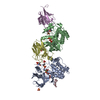
| ||||||||
|---|---|---|---|---|---|---|---|---|---|
| 1 | 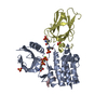
| ||||||||
| 2 | 
| ||||||||
| Unit cell |
|
-Components
-Protein , 2 types, 4 molecules ABDE
| #1: Protein | Mass: 30520.119 Da / Num. of mol.: 2 / Mutation: L33E Source method: isolated from a genetically manipulated source Source: (gene. exp.) Mycobacterium tuberculosis (strain ATCC 25618 / H37Rv) (bacteria)Strain: ATCC 25618 / H37Rv / Gene: pknB, Rv0014c, MTCY10H4.14c / Production host: References: UniProt: P9WI81, non-specific serine/threonine protein kinase #2: Protein | Mass: 17322.875 Da / Num. of mol.: 2 Source method: isolated from a genetically manipulated source Source: (gene. exp.) Mycobacterium tuberculosis (strain ATCC 25618 / H37Rv) (bacteria)Strain: ATCC 25618 / H37Rv / Gene: garA, Rv1827, MTCY1A11.16c / Production host: |
|---|
-Protein/peptide , 1 types, 1 molecules C
| #3: Protein/peptide | Mass: 443.539 Da / Num. of mol.: 1 Source method: isolated from a genetically manipulated source Source: (gene. exp.) Mycobacterium tuberculosis (strain ATCC 25618 / H37Rv) (bacteria)Production host: |
|---|
-Non-polymers , 4 types, 235 molecules 


| #4: Chemical |  Mass: 505.208 Da / Num. of mol.: 2 / Source method: obtained synthetically / Formula: C11H18N5O12P3 / Comment: AMP-PCP, energy-carrying molecule analogue*YM Mass: 505.208 Da / Num. of mol.: 2 / Source method: obtained synthetically / Formula: C11H18N5O12P3 / Comment: AMP-PCP, energy-carrying molecule analogue*YM#5: Chemical |  Mass: 24.305 Da / Num. of mol.: 2 / Source method: obtained synthetically / Formula: Mg Mass: 24.305 Da / Num. of mol.: 2 / Source method: obtained synthetically / Formula: Mg#6: Chemical |  Mass: 96.063 Da / Num. of mol.: 3 / Source method: obtained synthetically / Formula: SO4 Mass: 96.063 Da / Num. of mol.: 3 / Source method: obtained synthetically / Formula: SO4#7: Water | ChemComp-HOH / | Mass: 18.015 Da / Num. of mol.: 228 / Source method: isolated from a natural source / Formula: H2O |
|---|
-Details
| Has protein modification | Y |
|---|
-Experimental details
-Experiment
| Experiment | Method: X-RAY DIFFRACTION / Number of used crystals: 1 |
|---|
- Sample preparation
| Crystal | Density Matthews: 2.41 Å3/Da / Density % sol: 48.89 % |
|---|---|
| Crystal grow | Temperature: 291 K / Method: vapor diffusion, hanging drop Details: 100 mM Hepes-Na pH 7.5, 4 % w/v PEG 400, 2 M (NH4)2SO4, 4 mM AMP-PCP |
-Data collection
| Diffraction | Mean temperature: 100 K / Serial crystal experiment: N |
|---|---|
| Diffraction source | Source: SYNCHROTRON / Site: SOLEIL  / Beamline: PROXIMA 1 / Wavelength: 0.9763 Å / Beamline: PROXIMA 1 / Wavelength: 0.9763 Å |
| Detector | Type: ADSC QUANTUM 315r / Detector: CCD / Date: Dec 19, 2009 |
| Radiation | Protocol: SINGLE WAVELENGTH / Monochromatic (M) / Laue (L): M / Scattering type: x-ray |
| Radiation wavelength | Wavelength: 0.9763 Å / Relative weight: 1 |
| Reflection | Resolution: 2.37→38.85 Å / Num. obs: 36716 / % possible obs: 96.3 % / Redundancy: 4.6 % / Biso Wilson estimate: 67.39 Å2 / Rmerge(I) obs: 0.081 / Rpim(I) all: 0.04 / Net I/σ(I): 10.5 |
| Reflection shell | Resolution: 2.37→2.46 Å / Rmerge(I) obs: 0.439 / Rpim(I) all: 0.235 |
- Processing
| Software |
| ||||||||||||||||||||||||||||||||||||||||||||||||||||||||||||||||||||||||||||||||||||||||||||||||||||||||||||||||||||||||||||||||||||||||||||||||||||||||||||||||||||||||||||||||||||||||||||||||||||||||
|---|---|---|---|---|---|---|---|---|---|---|---|---|---|---|---|---|---|---|---|---|---|---|---|---|---|---|---|---|---|---|---|---|---|---|---|---|---|---|---|---|---|---|---|---|---|---|---|---|---|---|---|---|---|---|---|---|---|---|---|---|---|---|---|---|---|---|---|---|---|---|---|---|---|---|---|---|---|---|---|---|---|---|---|---|---|---|---|---|---|---|---|---|---|---|---|---|---|---|---|---|---|---|---|---|---|---|---|---|---|---|---|---|---|---|---|---|---|---|---|---|---|---|---|---|---|---|---|---|---|---|---|---|---|---|---|---|---|---|---|---|---|---|---|---|---|---|---|---|---|---|---|---|---|---|---|---|---|---|---|---|---|---|---|---|---|---|---|---|---|---|---|---|---|---|---|---|---|---|---|---|---|---|---|---|---|---|---|---|---|---|---|---|---|---|---|---|---|---|---|---|---|
| Refinement | Method to determine structure: MOLECULAR REPLACEMENT Starting model: 1O6Y Resolution: 2.37→38.85 Å / Cor.coef. Fo:Fc: 0.93 / Cor.coef. Fo:Fc free: 0.922 / SU R Cruickshank DPI: 0.364 / Cross valid method: THROUGHOUT / σ(F): 0 / SU R Blow DPI: 0.378 / SU Rfree Blow DPI: 0.227 / SU Rfree Cruickshank DPI: 0.227
| ||||||||||||||||||||||||||||||||||||||||||||||||||||||||||||||||||||||||||||||||||||||||||||||||||||||||||||||||||||||||||||||||||||||||||||||||||||||||||||||||||||||||||||||||||||||||||||||||||||||||
| Displacement parameters | Biso mean: 67.42 Å2
| ||||||||||||||||||||||||||||||||||||||||||||||||||||||||||||||||||||||||||||||||||||||||||||||||||||||||||||||||||||||||||||||||||||||||||||||||||||||||||||||||||||||||||||||||||||||||||||||||||||||||
| Refine analyze | Luzzati coordinate error obs: 0.36 Å | ||||||||||||||||||||||||||||||||||||||||||||||||||||||||||||||||||||||||||||||||||||||||||||||||||||||||||||||||||||||||||||||||||||||||||||||||||||||||||||||||||||||||||||||||||||||||||||||||||||||||
| Refinement step | Cycle: 1 / Resolution: 2.37→38.85 Å
| ||||||||||||||||||||||||||||||||||||||||||||||||||||||||||||||||||||||||||||||||||||||||||||||||||||||||||||||||||||||||||||||||||||||||||||||||||||||||||||||||||||||||||||||||||||||||||||||||||||||||
| Refine LS restraints |
| ||||||||||||||||||||||||||||||||||||||||||||||||||||||||||||||||||||||||||||||||||||||||||||||||||||||||||||||||||||||||||||||||||||||||||||||||||||||||||||||||||||||||||||||||||||||||||||||||||||||||
| LS refinement shell | Resolution: 2.37→2.44 Å / Total num. of bins used: 18
| ||||||||||||||||||||||||||||||||||||||||||||||||||||||||||||||||||||||||||||||||||||||||||||||||||||||||||||||||||||||||||||||||||||||||||||||||||||||||||||||||||||||||||||||||||||||||||||||||||||||||
| Refinement TLS params. | Method: refined / Refine-ID: X-RAY DIFFRACTION
| ||||||||||||||||||||||||||||||||||||||||||||||||||||||||||||||||||||||||||||||||||||||||||||||||||||||||||||||||||||||||||||||||||||||||||||||||||||||||||||||||||||||||||||||||||||||||||||||||||||||||
| Refinement TLS group |
|
