 Movie
Movie Controller

[English] 日本語
 Yorodumi
- PDB-6i2q: Crystal structure of the wild-type SucA domain of Mycobacterium s... -
+ Open data

- Basic information
| Entry | Database: PDB / ID: 6i2q | |||||||||
|---|---|---|---|---|---|---|---|---|---|---|
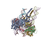
| Title | Crystal structure of the wild-type SucA domain of Mycobacterium smegmatis KGD (alpha-ketoglutarate decarboxylase), in complex with GarA | |||||||||
 Components Components |
| |||||||||
 Keywords Keywords | OXIDOREDUCTASE / oxoglutarate dehydrogenase / decarboxylase | |||||||||
| Function / homology |  Function and homology information Function and homology information2-hydroxy-3-oxoadipate synthase / 2-oxoglutarate decarboxylase / 2-oxoglutarate decarboxylase activity / 2-hydroxy-3-oxoadipate synthase activity / oxoglutarate dehydrogenase (succinyl-transferring) / oxoglutarate dehydrogenase (succinyl-transferring) activity / dihydrolipoyllysine-residue succinyltransferase / dihydrolipoyllysine-residue succinyltransferase activity / oxoglutarate dehydrogenase complex / thiamine pyrophosphate binding ...2-hydroxy-3-oxoadipate synthase / 2-oxoglutarate decarboxylase / 2-oxoglutarate decarboxylase activity / 2-hydroxy-3-oxoadipate synthase activity / oxoglutarate dehydrogenase (succinyl-transferring) / oxoglutarate dehydrogenase (succinyl-transferring) activity / dihydrolipoyllysine-residue succinyltransferase / dihydrolipoyllysine-residue succinyltransferase activity / oxoglutarate dehydrogenase complex / thiamine pyrophosphate binding / tricarboxylic acid cycle / magnesium ion binding / cytosol Similarity search - Function | |||||||||
| Biological species |  Mycobacterium smegmatis (bacteria) Mycobacterium smegmatis (bacteria) | |||||||||
| Method |  X-RAY DIFFRACTION / SYNCHROTRON / MOLECULAR REPLACEMENT / Resolution: 2.15 Å X-RAY DIFFRACTION / SYNCHROTRON / MOLECULAR REPLACEMENT / Resolution: 2.15 Å | |||||||||
 Authors Authors | Wagner, T. / Bellinzoni, M. / Alzari, P.M. | |||||||||
 Citation Citation | Journal: Sci.Signal. / Year: 2019 Title: Structural insights into the functional versatility of an FHA domain protein in mycobacterial signaling. Authors: Wagner, T. / Andre-Leroux, G. / Hindie, V. / Barilone, N. / Lisa, M.N. / Hoos, S. / Raynal, B. / Vulliez-Le Normand, B. / O'Hare, H.M. / Bellinzoni, M. / Alzari, P.M. | |||||||||
| History |
|
- Structure visualization
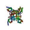
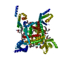
| Structure viewer | Molecule: MolmilJmol/JSmol |
|---|
- Downloads & links
-Download
| PDBx/mmCIF format | 6i2q.cif.gz | 389.6 KB | Display | PDBx/mmCIF format |
|---|---|---|---|---|
| PDB format | pdb6i2q.ent.gz | 309.3 KB | Display | PDB format |
| PDBx/mmJSON format | 6i2q.json.gz | Tree view | PDBx/mmJSON format | |
| Others |  Other downloads Other downloads |
-Validation report
| Arichive directory | https://data.pdbj.org/pub/pdb/validation_reports/i2/6i2qftp://data.pdbj.org/pub/pdb/validation_reports/i2/6i2q | HTTPS FTP |
|---|
-Related structure data
| Related structure data | 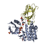 6i2pC  6i2rC  6i2sC 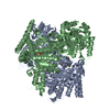 2yicS S: Starting model for refinement C: citing same article ( |
|---|---|
| Similar structure data |
-Links
 PDBj
PDBj


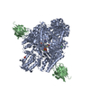
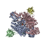
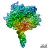
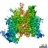
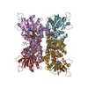
- Assembly
| Deposited unit | 
| ||||||||
|---|---|---|---|---|---|---|---|---|---|
| 1 | 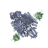
| ||||||||
| Unit cell |
|
-Components
-Protein , 2 types, 2 molecules AB
| #1: Protein | Mass: 97166.648 Da / Num. of mol.: 1 Source method: isolated from a genetically manipulated source Source: (gene. exp.) Mycobacterium smegmatis (strain ATCC 700084 / mc(2)155) (bacteria)Strain: ATCC 700084 / mc(2)155 / Gene: kgd, sucA, MSMEG_5049, MSMEI_4922 / Production host: References: UniProt: A0R2B1, 2-hydroxy-3-oxoadipate synthase, 2-oxoglutarate decarboxylase, oxoglutarate dehydrogenase (succinyl-transferring), dihydrolipoyllysine-residue succinyltransferase |
|---|---|
| #2: Protein | Mass: 12112.292 Da / Num. of mol.: 1 Source method: isolated from a genetically manipulated source Source: (gene. exp.) Mycobacterium smegmatis (strain ATCC 700084 / mc(2)155) (bacteria)Strain: ATCC 700084 / mc(2)155 / Gene: garA, MSMEG_3647, MSMEI_3561 / Production host: |
-Non-polymers , 4 types, 359 molecules 


| #3: Chemical | ChemComp-TPP /  Mass: 425.314 Da / Num. of mol.: 1 / Source method: obtained synthetically / Formula: C12H19N4O7P2S Mass: 425.314 Da / Num. of mol.: 1 / Source method: obtained synthetically / Formula: C12H19N4O7P2S |
|---|---|
| #4: Chemical | ChemComp-MG /  Mass: 24.305 Da / Num. of mol.: 1 / Source method: obtained synthetically / Formula: Mg Mass: 24.305 Da / Num. of mol.: 1 / Source method: obtained synthetically / Formula: Mg |
| #5: Chemical | ChemComp-CA /  Mass: 40.078 Da / Num. of mol.: 1 / Source method: obtained synthetically / Formula: Ca Mass: 40.078 Da / Num. of mol.: 1 / Source method: obtained synthetically / Formula: Ca |
| #6: Water | ChemComp-HOH / Mass: 18.015 Da / Num. of mol.: 356 / Source method: isolated from a natural source / Formula: H2O |
-Experimental details
-Experiment
| Experiment | Method: X-RAY DIFFRACTION / Number of used crystals: 1 |
|---|
- Sample preparation
| Crystal | Density Matthews: 2.79 Å3/Da / Density % sol: 55.88 % |
|---|---|
| Crystal grow | Temperature: 291 K / Method: vapor diffusion, hanging drop / Details: 100 mM Hepes-Na pH 7.0, 50% MPD |
-Data collection
| Diffraction | Mean temperature: 100 K / Serial crystal experiment: N |
|---|---|
| Diffraction source | Source: SYNCHROTRON / Site: SOLEIL  / Beamline: PROXIMA 1 / Wavelength: 0.98 Å / Beamline: PROXIMA 1 / Wavelength: 0.98 Å |
| Detector | Type: ADSC QUANTUM 315r / Detector: CCD / Date: May 29, 2009 |
| Radiation | Protocol: SINGLE WAVELENGTH / Monochromatic (M) / Laue (L): M / Scattering type: x-ray |
| Radiation wavelength | Wavelength: 0.98 Å / Relative weight: 1 |
| Reflection | Resolution: 2.15→47.83 Å / Num. obs: 65695 / % possible obs: 98.9 % / Redundancy: 5 % / Biso Wilson estimate: 44.27 Å2 / Rmerge(I) obs: 0.062 / Rpim(I) all: 0.03 / Net I/σ(I): 16.6 |
| Reflection shell | Resolution: 2.15→2.2 Å / Rmerge(I) obs: 0.747 / Rpim(I) all: 0.361 |
- Processing
| Software |
| ||||||||||||||||||||||||||||||||||||||||||||||||||||||||||||||||||||||||||||||||||||||||||||||||||||||||||||||||||
|---|---|---|---|---|---|---|---|---|---|---|---|---|---|---|---|---|---|---|---|---|---|---|---|---|---|---|---|---|---|---|---|---|---|---|---|---|---|---|---|---|---|---|---|---|---|---|---|---|---|---|---|---|---|---|---|---|---|---|---|---|---|---|---|---|---|---|---|---|---|---|---|---|---|---|---|---|---|---|---|---|---|---|---|---|---|---|---|---|---|---|---|---|---|---|---|---|---|---|---|---|---|---|---|---|---|---|---|---|---|---|---|---|---|---|---|
| Refinement | Method to determine structure: MOLECULAR REPLACEMENT Starting model: 2YIC Resolution: 2.15→31.48 Å / Cor.coef. Fo:Fc: 0.96 / Cor.coef. Fo:Fc free: 0.953 / SU R Cruickshank DPI: 0.162 / Cross valid method: THROUGHOUT / σ(F): 0 / SU R Blow DPI: 0.163 / SU Rfree Blow DPI: 0.139 / SU Rfree Cruickshank DPI: 0.14
| ||||||||||||||||||||||||||||||||||||||||||||||||||||||||||||||||||||||||||||||||||||||||||||||||||||||||||||||||||
| Displacement parameters | Biso mean: 56.3 Å2
| ||||||||||||||||||||||||||||||||||||||||||||||||||||||||||||||||||||||||||||||||||||||||||||||||||||||||||||||||||
| Refine analyze | Luzzati coordinate error obs: 0.22 Å | ||||||||||||||||||||||||||||||||||||||||||||||||||||||||||||||||||||||||||||||||||||||||||||||||||||||||||||||||||
| Refinement step | Cycle: 1 / Resolution: 2.15→31.48 Å
| ||||||||||||||||||||||||||||||||||||||||||||||||||||||||||||||||||||||||||||||||||||||||||||||||||||||||||||||||||
| Refine LS restraints |
| ||||||||||||||||||||||||||||||||||||||||||||||||||||||||||||||||||||||||||||||||||||||||||||||||||||||||||||||||||
| LS refinement shell | Resolution: 2.15→2.21 Å / Total num. of bins used: 20
| ||||||||||||||||||||||||||||||||||||||||||||||||||||||||||||||||||||||||||||||||||||||||||||||||||||||||||||||||||
| Refinement TLS params. | Method: refined / Refine-ID: X-RAY DIFFRACTION
| ||||||||||||||||||||||||||||||||||||||||||||||||||||||||||||||||||||||||||||||||||||||||||||||||||||||||||||||||||
| Refinement TLS group |
|
