 Movie
Movie Controller


+ Open data

- Basic information
| Entry | Database: PDB / ID: 6pe2 | |||||||||
|---|---|---|---|---|---|---|---|---|---|---|
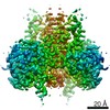
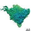
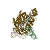
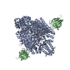
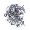
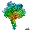
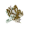
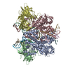
| Title | Drosophila P element transposase strand transfer complex | |||||||||
 Components Components |
| |||||||||
 Keywords Keywords | TRANSFERASE/DNA / transposase / Strand Transfer Complex / TRANSFERASE-DNA / TRANSFERASE-DNA complex | |||||||||
| Function / homology |  Function and homology information Function and homology informationP-element binding / transposase activity / DNA transposition / DNA integration / Transferases; Transferring phosphorus-containing groups; Nucleotidyltransferases / transferase activity / zinc ion binding Similarity search - Function | |||||||||
| Biological species |  | |||||||||
| Method | ELECTRON MICROSCOPY / single particle reconstruction / cryo EM / Resolution: 4 Å | |||||||||
 Authors Authors | Kellogg, E.H. / Nogales, E. / Ghanim, G. / Rio, D.C. | |||||||||
| Funding support |  United States, 2items United States, 2items
| |||||||||
 Citation Citation | Journal: Nat Struct Mol Biol / Year: 2019 Title: Structure of a P element transposase-DNA complex reveals unusual DNA structures and GTP-DNA contacts. Authors: George E Ghanim / Elizabeth H Kellogg / Eva Nogales / Donald C Rio / Abstract: P element transposase catalyzes the mobility of P element DNA transposons within the Drosophila genome. P element transposase exhibits several unique properties, including the requirement for a ...P element transposase catalyzes the mobility of P element DNA transposons within the Drosophila genome. P element transposase exhibits several unique properties, including the requirement for a guanosine triphosphate cofactor and the generation of long staggered DNA breaks during transposition. To gain insights into these features, we determined the atomic structure of the Drosophila P element transposase strand transfer complex using cryo-EM. The structure of this post-transposition nucleoprotein complex reveals that the terminal single-stranded transposon DNA adopts unusual A-form and distorted B-form helical geometries that are stabilized by extensive protein-DNA interactions. Additionally, we infer that the bound guanosine triphosphate cofactor interacts with the terminal base of the transposon DNA, apparently to position the P element DNA for catalysis. Our structure provides the first view of the P element transposase superfamily, offers new insights into P element transposition and implies a transposition pathway fundamentally distinct from other cut-and-paste DNA transposases. | |||||||||
| History |
|
- Structure visualization
| Movie |
Movie viewer |
|---|---|
| Structure viewer | Molecule: MolmilJmol/JSmol |
- Downloads & links
-Download
| PDBx/mmCIF format | 6pe2.cif.gz | 303 KB | Display | PDBx/mmCIF format |
|---|---|---|---|---|
| PDB format | pdb6pe2.ent.gz | 231.4 KB | Display | PDB format |
| PDBx/mmJSON format | 6pe2.json.gz | Tree view | PDBx/mmJSON format | |
| Others |  Other downloads Other downloads |
-Validation report
| Arichive directory | https://data.pdbj.org/pub/pdb/validation_reports/pe/6pe2ftp://data.pdbj.org/pub/pdb/validation_reports/pe/6pe2 | HTTPS FTP |
|---|
-Related structure data
| Related structure data | 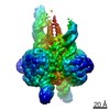 20321MC 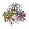 6p5aC C: citing same article ( M: map data used to model this data |
|---|---|
| Similar structure data |
-Links
 PDBj
PDBj


- Assembly
| Deposited unit | 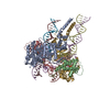
|
|---|---|
| 1 |
|
-Components
-Transposable element P ... , 2 types, 4 molecules AGBH
| #1: Protein | Mass: 65525.992 Da / Num. of mol.: 2 / Fragment: N-terminal domain (UNP residues 1-569) Source method: isolated from a genetically manipulated source Source: (gene. exp.)  Spodoptera frugiperda (fall armyworm) Spodoptera frugiperda (fall armyworm)References: UniProt: Q7M3K2, Transferases; Transferring phosphorus-containing groups; Nucleotidyltransferases #2: Protein | Mass: 16086.797 Da / Num. of mol.: 2 / Fragment: C-terminal domain (UNP residues 613-747) Source method: isolated from a genetically manipulated source Source: (gene. exp.) Spodoptera frugiperda (fall armyworm)References: UniProt: Q7M3K2, Transferases; Transferring phosphorus-containing groups; Nucleotidyltransferases |
|---|
-DNA chain , 3 types, 6 molecules CIDJEK
| #3: DNA chain | Mass: 4827.157 Da / Num. of mol.: 2 / Source method: obtained synthetically / Source: (synth.) #4: DNA chain | Mass: 11704.529 Da / Num. of mol.: 2 / Source method: obtained synthetically / Source: (synth.) #5: DNA chain | Mass: 24400.576 Da / Num. of mol.: 2 / Source method: obtained synthetically / Source: (synth.) |
|---|
-Non-polymers , 2 types, 6 molecules 

| #6: Chemical |  Mass: 523.180 Da / Num. of mol.: 2 / Source method: obtained synthetically / Formula: C10H16N5O14P3 / Feature type: SUBJECT OF INVESTIGATION / Comment: GTP, energy-carrying molecule*YM Mass: 523.180 Da / Num. of mol.: 2 / Source method: obtained synthetically / Formula: C10H16N5O14P3 / Feature type: SUBJECT OF INVESTIGATION / Comment: GTP, energy-carrying molecule*YM#7: Chemical | ChemComp-MG /  Mass: 24.305 Da / Num. of mol.: 4 / Source method: obtained synthetically / Formula: Mg Mass: 24.305 Da / Num. of mol.: 4 / Source method: obtained synthetically / Formula: Mg |
|---|
-Details
| Has ligand of interest | Y |
|---|
-Experimental details
-Experiment
| Experiment | Method: ELECTRON MICROSCOPY |
|---|---|
| EM experiment | Aggregation state: PARTICLE / 3D reconstruction method: single particle reconstruction |
- Sample preparation
| Component | Name: Ternary complex of P element transposase in complex with strand transfer DNA Type: COMPLEX / Details: C1 reconstruction / Entity ID: #1-#5 / Source: RECOMBINANT | ||||||||||||||||||||||||||||||
|---|---|---|---|---|---|---|---|---|---|---|---|---|---|---|---|---|---|---|---|---|---|---|---|---|---|---|---|---|---|---|---|
| Molecular weight | Experimental value: NO | ||||||||||||||||||||||||||||||
| Source (natural) | Organism: | ||||||||||||||||||||||||||||||
| Source (recombinant) | Organism: Spodoptera frugiperda (fall armyworm) | ||||||||||||||||||||||||||||||
| Buffer solution | pH: 7.6 | ||||||||||||||||||||||||||||||
| Buffer component |
| ||||||||||||||||||||||||||||||
| Specimen | Embedding applied: NO / Shadowing applied: NO / Staining applied: NO / Vitrification applied: YES | ||||||||||||||||||||||||||||||
| Specimen support | Details: unspecified | ||||||||||||||||||||||||||||||
| Vitrification | Instrument: FEI VITROBOT MARK IV / Cryogen name: ETHANE / Humidity: 100 % / Chamber temperature: 277 K Details: 4 microliters of concentrated STC complex was applied to a Quantifoil 1.2/1.3 UltraAuFoil grid. After a 30 second incubation, the sample was blotted using blot force of 8 pN and a blot time of 6 seconds. |
- Electron microscopy imaging
| Experimental equipment |  Model: Talos Arctica / Image courtesy: FEI Company |
|---|---|
| Microscopy | Model: FEI TECNAI ARCTICA / Details: dataset collected at 40 degree tilt |
| Electron gun | Electron source:  FIELD EMISSION GUN / Accelerating voltage: 200 kV / Illumination mode: FLOOD BEAM FIELD EMISSION GUN / Accelerating voltage: 200 kV / Illumination mode: FLOOD BEAM |
| Electron lens | Mode: BRIGHT FIELD / Nominal defocus max: 3000 nm / Nominal defocus min: 1000 nm / Cs: 2.7 mm / C2 aperture diameter: 100 µm / Alignment procedure: COMA FREE |
| Specimen holder | Cryogen: NITROGEN |
| Image recording | Average exposure time: 10 sec. / Electron dose: 60 e/Å2 / Detector mode: SUPER-RESOLUTION / Film or detector model: GATAN K2 SUMMIT (4k x 4k) / Num. of grids imaged: 1 / Num. of real images: 1857 |
| EM imaging optics | Spherical aberration corrector: none |
| Image scans | Width: 7420 / Height: 7676 / Movie frames/image: 39 |
- Processing
| EM software |
| ||||||||||||||||||||||||||||||||||||||||
|---|---|---|---|---|---|---|---|---|---|---|---|---|---|---|---|---|---|---|---|---|---|---|---|---|---|---|---|---|---|---|---|---|---|---|---|---|---|---|---|---|---|
| CTF correction | Type: PHASE FLIPPING ONLY | ||||||||||||||||||||||||||||||||||||||||
| Symmetry | Point symmetry: C1 (asymmetric) | ||||||||||||||||||||||||||||||||||||||||
| 3D reconstruction | Resolution: 4 Å / Resolution method: FSC 0.143 CUT-OFF / Num. of particles: 253209 / Algorithm: FOURIER SPACE / Symmetry type: POINT | ||||||||||||||||||||||||||||||||||||||||
| Atomic model building | B value: 150 / Protocol: AB INITIO MODEL / Space: REAL / Target criteria: correlation coefficient |
