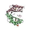
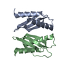
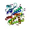
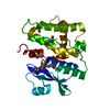
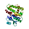
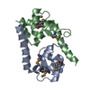
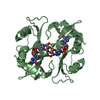
[English] 日本語
Yorodumi
- PDB-6gp6: MamM CTD - Copper form -

+
Open data


ID or keywords:

Loading...

-
Basic information

Entry
Database: PDB / ID: 6gp6
TitleMamM CTD - Copper form
ComponentsMagnetosome protein MamM
KeywordsMETAL TRANSPORT / Cation diffusion facilitator / magnetotactic bacteria
Function / homology
Function and homology information


magnetosome membrane / monoatomic cation transmembrane transporter activity / iron ion transport / metal ion binding / plasma membrane
Similarity search - Function
: / : / Cation efflux protein, cytoplasmic domain / Dimerisation domain of Zinc Transporter / Cation efflux protein, cytoplasmic domain superfamily / Cation efflux protein / Cation efflux transmembrane domain superfamily / Cation efflux family
Similarity search - Domain/homology
BETA-MERCAPTOETHANOL / COPPER (II) ION / Magnetosome protein MamM / Magnetosome protein MamM
Similarity search - Component
Biological speciesMagnetospirillum gryphiswaldense MSR-1 (magnetotactic)
MethodX-RAY DIFFRACTION / SYNCHROTRON / MOLECULAR REPLACEMENT / Resolution: 2.146 Å
AuthorsBarber-Zucker, S. / Zarivach, R.
Funding support Israel, 2items
OrganizationGrant numberCountry
Israel Science Foundation 167/16 Israel
Israel Ministry of Science, Technology and Space Israel
CitationJournal: To Be Published
Title: MamM CTD - Copper form
Authors: Barber-Zucker, S. / Zarivach, R.
History
DepositionJun 5, 2018Deposition site: PDBE / Processing site: PDBE
Revision 1.0Jun 19, 2019Provider: repository / Type: Initial release
Revision 1.1Jan 17, 2024Group: Data collection / Database references ...Data collection / Database references / Derived calculations / Refinement description
Category: chem_comp_atom / chem_comp_bond ...chem_comp_atom / chem_comp_bond / database_2 / pdbx_initial_refinement_model / struct_conn / struct_ncs_dom_lim
Item: _database_2.pdbx_DOI / _database_2.pdbx_database_accession ..._database_2.pdbx_DOI / _database_2.pdbx_database_accession / _struct_conn.pdbx_dist_value / _struct_conn.ptnr1_auth_asym_id / _struct_conn.ptnr1_auth_comp_id / _struct_conn.ptnr1_auth_seq_id / _struct_conn.ptnr1_label_asym_id / _struct_conn.ptnr1_label_atom_id / _struct_conn.ptnr1_label_comp_id / _struct_conn.ptnr1_label_seq_id / _struct_conn.ptnr2_auth_asym_id / _struct_conn.ptnr2_auth_comp_id / _struct_conn.ptnr2_auth_seq_id / _struct_conn.ptnr2_label_asym_id / _struct_conn.ptnr2_label_atom_id / _struct_conn.ptnr2_label_comp_id / _struct_conn.ptnr2_label_seq_id / _struct_ncs_dom_lim.beg_auth_comp_id / _struct_ncs_dom_lim.beg_label_asym_id / _struct_ncs_dom_lim.beg_label_comp_id / _struct_ncs_dom_lim.beg_label_seq_id / _struct_ncs_dom_lim.end_auth_comp_id / _struct_ncs_dom_lim.end_label_asym_id / _struct_ncs_dom_lim.end_label_comp_id / _struct_ncs_dom_lim.end_label_seq_id

-
Structure visualization

Structure viewerMolecule:
MolmilJmol/JSmol

Downloads & links

-
Assembly

Deposited unit
A: Magnetosome protein MamM
B: Magnetosome protein MamM
hetero molecules


Theoretical massNumber of molelcules
Total (without water)24,0906
Polymers23,8212
Non-polymers2694
Water37821
1


  • Idetical with deposited unit
  • defined by author&software
  • Evidence: gel filtration
TypeNameSymmetry operationNumber
identity operation1_555x,y,z1
Buried area2090 Å2
ΔGint-36 kcal/mol
Surface area10590 Å2
MethodPISA
Unit cell
Length a, b, c (Å)28.930, 73.750, 89.410
Angle α, β, γ (deg.)90.000, 90.000, 90.000
Int Tables number18
Space group name H-MP22121
Noncrystallographic symmetry (NCS)NCS domain:
IDEns-IDDetails
11(chain A and (resid 212 through 237 or resid 239 through 258 or resid 260 through 301))
21(chain B and (resid 212 through 237 or resid 239 through 258 or resid 260 through 301))

NCS domain segments:

Ens-ID: 1

Dom-IDComponent-IDBeg auth comp-IDBeg label comp-IDEnd auth comp-IDEnd label comp-IDSelection detailsAuth asym-IDLabel asym-IDAuth seq-IDLabel seq-ID
11SERSERLEULEU(chain A and (resid 212 through 237 or resid 239 through 258 or resid 260 through 301))AA212 - 2372 - 27
12ALAALAASNASN(chain A and (resid 212 through 237 or resid 239 through 258 or resid 260 through 301))AA239 - 25829 - 48
13VALVALPHEPHE(chain A and (resid 212 through 237 or resid 239 through 258 or resid 260 through 301))AA260 - 30150 - 91
21SERSERLEULEU(chain B and (resid 212 through 237 or resid 239 through 258 or resid 260 through 301))BB212 - 2372 - 27
22ALAALAASNASN(chain B and (resid 212 through 237 or resid 239 through 258 or resid 260 through 301))BB239 - 25829 - 48
23VALVALPHEPHE(chain B and (resid 212 through 237 or resid 239 through 258 or resid 260 through 301))BB260 - 30150 - 91

-
Components

#1: Protein Magnetosome protein MamM / Magnetosome protein MamM / Cation efflux protein family / MamM protein


Mass: 11910.398 Da / Num. of mol.: 2
Source method: isolated from a genetically manipulated source
Source: (gene. exp.) Magnetospirillum gryphiswaldense MSR-1 (magnetotactic)
Gene: mamM, mgI491, MGR_4095 / Production host: Escherichia coli BL21(DE3) (bacteria) / Variant (production host): Rosetta / References: UniProt: Q6NE57, UniProt: V6F235*PLUS
#2: Chemical ChemComp-CU / COPPER (II) ION


Mass: 63.546 Da / Num. of mol.: 3
Source method: isolated from a genetically manipulated source
Formula: Cu / Feature type: SUBJECT OF INVESTIGATION
#3: Chemical ChemComp-BME / BETA-MERCAPTOETHANOL


Mass: 78.133 Da / Num. of mol.: 1 / Source method: obtained synthetically / Formula: C2H6OS
#4: Water ChemComp-HOH / water


Mass: 18.015 Da / Num. of mol.: 21 / Source method: isolated from a natural source / Formula: H2O

-
Experimental details

-
Experiment

ExperimentMethod: X-RAY DIFFRACTION / Number of used crystals: 1

-
Sample preparation

CrystalDensity Matthews: 2.09 Å3/Da / Density % sol: 41.26 %
Crystal growTemperature: 293 K / Method: vapor diffusion, sitting drop
Details: 0.2M NaCl, 0.1M Tris pH=8.7, 25% PEG 3350, 1.7 mM CuSO4

-
Data collection

DiffractionMean temperature: 100 K
Diffraction sourceSource: SYNCHROTRON / Site: ESRF / Beamline: MASSIF-3 / Wavelength: 0.96771 Å
DetectorType: DECTRIS EIGER X 4M / Detector: PIXEL / Date: Feb 23, 2017
RadiationProtocol: SINGLE WAVELENGTH / Monochromatic (M) / Laue (L): M / Scattering type: x-ray
Radiation wavelengthWavelength: 0.96771 Å / Relative weight: 1
ReflectionResolution: 2.14→44.71 Å / Num. obs: 11127 / % possible obs: 99.6 % / Redundancy: 5.8 % / Biso Wilson estimate: 31.35 Å2 / CC1/2: 0.996 / Rmerge(I) obs: 0.106 / Rpim(I) all: 0.049 / Rrim(I) all: 0.118 / Net I/σ(I): 8.5 / Num. measured all: 64227
Reflection shell

Diffraction-ID: 1

Resolution (Å)Redundancy (%)Rmerge(I) obsNum. measured allNum. unique obsCC1/2Rpim(I) allRrim(I) allNet I/σ(I) obs% possible all
2.14-2.25.81.86350428640.4360.8282.044196.9
9.08-44.714.60.0498411840.9980.0240.05522.198.1

-
Processing

Software
NameClassification
PHENIXrefinement
XDSdata reduction
Aimlessdata scaling
PDB_EXTRACTdata extraction
PHASERphasing
RefinementMethod to determine structure: MOLECULAR REPLACEMENT
Starting model: 3W5X
Resolution: 2.146→44.705 Å / SU ML: 0.35 / Cross valid method: THROUGHOUT / σ(F): 1.35 / Phase error: 35.39
RfactorNum. reflection% reflection
Rfree0.2751 419 4.7 %
Rwork0.2201 --
obs0.2231 8919 80.67 %
Solvent computationShrinkage radii: 0.9 Å / VDW probe radii: 1.11 Å
Displacement parametersBiso max: 144.3 Å2 / Biso mean: 43.6275 Å2 / Biso min: 17.88 Å2
Refinement stepCycle: final / Resolution: 2.146→44.705 Å
ProteinNucleic acidLigandSolventTotal
Num. atoms1495 0 7 21 1523
Biso mean--62.01 41.44 -
Num. residues----194
Refine LS restraints
Refine-IDTypeDev idealNumber
X-RAY DIFFRACTIONf_bond_d0.0091537
X-RAY DIFFRACTIONf_angle_d1.0762081
X-RAY DIFFRACTIONf_chiral_restr0.065237
X-RAY DIFFRACTIONf_plane_restr0.007278
X-RAY DIFFRACTIONf_dihedral_angle_d14.6621103
Refine LS restraints NCS
Ens-IDDom-IDAuth asym-IDNumberRefine-IDRmsType
11A751X-RAY DIFFRACTION15.82TORSIONAL
12B751X-RAY DIFFRACTION15.82TORSIONAL
LS refinement shell

Refine-ID: X-RAY DIFFRACTION / Rfactor Rfree error: 0 / Total num. of bins used: 3

Resolution (Å)Rfactor RfreeNum. reflection RfreeRfactor RworkNum. reflection RworkNum. reflection all% reflection obs (%)
2.1465-2.45710.3451710.25611486155743
2.4571-3.09560.31821670.2683384355198
3.0956-44.71470.25141810.195736303811100
Refinement TLS params.Method: refined / Origin x: -1.6872 Å / Origin y: -15.8303 Å / Origin z: 34.726 Å
111213212223313233
T0.2509 Å20.0162 Å20.1285 Å2-0.1884 Å2-0.0177 Å2--0.2661 Å2
L0.8908 °2-0.1281 °20.2964 °2-2.4589 °2-0.4686 °2--2.5525 °2
S0.0593 Å °-0.1085 Å °0.1033 Å °0.2721 Å °0.0352 Å °0.0053 Å °-0.0215 Å °-0.1851 Å °-0.0706 Å °
Refinement TLS group
IDRefine-IDRefine TLS-IDSelection detailsAuth asym-IDAuth seq-ID
1X-RAY DIFFRACTION1allA212 - 314
2X-RAY DIFFRACTION1allB211 - 301
3X-RAY DIFFRACTION1allC2
4X-RAY DIFFRACTION1allC3 - 4
5X-RAY DIFFRACTION1allD1 - 21
6X-RAY DIFFRACTION1allE1

+
About Yorodumi

-
News

-
Feb 9, 2022. New format data for meta-information of EMDB entries

New format data for meta-information of EMDB entries

  • Version 3 of the EMDB header file is now the official format.
  • The previous official version 1.9 will be removed from the archive.

Related info.:EMDB header

External links:wwPDB to switch to version 3 of the EMDB data model

-
Aug 12, 2020. Covid-19 info

Covid-19 info

URL: https://pdbjlvh1.pdbj.org/emnavi/covid19.php

New page: Covid-19 featured information page in EM Navigator.

Related info.:Covid-19 info / Mar 5, 2020. Novel coronavirus structure data

+
Mar 5, 2020. Novel coronavirus structure data

Novel coronavirus structure data

Related info.:Yorodumi Speices / Aug 12, 2020. Covid-19 info

External links:COVID-19 featured content - PDBj / Molecule of the Month (242):Coronavirus Proteases

+
Jan 31, 2019. EMDB accession codes are about to change! (news from PDBe EMDB page)

EMDB accession codes are about to change! (news from PDBe EMDB page)

  • The allocation of 4 digits for EMDB accession codes will soon come to an end. Whilst these codes will remain in use, new EMDB accession codes will include an additional digit and will expand incrementally as the available range of codes is exhausted. The current 4-digit format prefixed with “EMD-” (i.e. EMD-XXXX) will advance to a 5-digit format (i.e. EMD-XXXXX), and so on. It is currently estimated that the 4-digit codes will be depleted around Spring 2019, at which point the 5-digit format will come into force.
  • The EM Navigator/Yorodumi systems omit the EMD- prefix.

Related info.:Q: What is EMD? / ID/Accession-code notation in Yorodumi/EM Navigator

External links:EMDB Accession Codes are Changing Soon! / Contact to PDBj

+
Jul 12, 2017. Major update of PDB

Major update of PDB

  • wwPDB released updated PDB data conforming to the new PDBx/mmCIF dictionary.
  • This is a major update changing the version number from 4 to 5, and with Remediation, in which all the entries are updated.
  • In this update, many items about electron microscopy experimental information are reorganized (e.g. em_software).
  • Now, EM Navigator and Yorodumi are based on the updated data.

External links:wwPDB Remediation / Enriched Model Files Conforming to OneDep Data Standards Now Available in the PDB FTP Archive

-
Yorodumi

Thousand views of thousand structures

  • Yorodumi is a browser for structure data from EMDB, PDB, SASBDB, etc.
  • This page is also the successor to EM Navigator detail page, and also detail information page/front-end page for Omokage search.
  • The word "yorodu" (or yorozu) is an old Japanese word meaning "ten thousand". "mi" (miru) is to see.

Related info.:EMDB / PDB / SASBDB / Comparison of 3 databanks / Yorodumi Search / Aug 31, 2016. New EM Navigator & Yorodumi / Yorodumi Papers / Jmol/JSmol / Function and homology information / Changes in new EM Navigator and Yorodumi

Read more