[English] 日本語
Yorodumi
- PDB-5elg: The structure of DHAR1 from Arabidopsis thaliana -

+
Open data


ID or keywords:

Loading...

-
Basic information

Entry
Database: PDB / ID: 5elg
TitleThe structure of DHAR1 from Arabidopsis thaliana
ComponentsGlutathione S-transferase DHAR1, mitochondrial
KeywordsTRANSFERASE / Dehydroascobate reductase / glutathione S-transferase / Arabidopsis thaliana / CLIC / Chloride ion channel
Function / homology
Function and homology information


response to symbiotic fungus / regulation of biological process involved in symbiotic interaction / acquisition of seed longevity / positive regulation of salicylic acid mediated signaling pathway / protein glutathionylation / ascorbate glutathione cycle / glutathione dehydrogenase (ascorbate) / glutathione dehydrogenase (ascorbate) activity / L-ascorbic acid metabolic process / response to jasmonic acid ...response to symbiotic fungus / regulation of biological process involved in symbiotic interaction / acquisition of seed longevity / positive regulation of salicylic acid mediated signaling pathway / protein glutathionylation / ascorbate glutathione cycle / glutathione dehydrogenase (ascorbate) / glutathione dehydrogenase (ascorbate) activity / L-ascorbic acid metabolic process / response to jasmonic acid / response to ozone / apoplast / plant-type vacuole / chloroplast stroma / monoatomic ion channel complex / response to zinc ion / glutathione transferase / glutathione transferase activity / monoatomic ion transmembrane transport / chloroplast / defense response / cellular response to hydrogen peroxide / peroxisome / copper ion binding / mitochondrion / nucleus / plasma membrane / cytosol
Similarity search - Function
Dehydroascorbate reductases DHAR1/2/3/4 / Glutathione S-transferase, N-terminal domain / Glutathione S-transferase, N-terminal domain / Glutathione S-transferase, C-terminal domain / Glutathione transferase family / Glutathione S-transferase Yfyf (Class Pi); Chain A, domain 2 - #10 / Glutathione S-transferase Yfyf (Class Pi); Chain A, domain 2 / Soluble glutathione S-transferase N-terminal domain profile. / Glutathione S-transferase, C-terminal-like / Soluble glutathione S-transferase C-terminal domain profile. ...Dehydroascorbate reductases DHAR1/2/3/4 / Glutathione S-transferase, N-terminal domain / Glutathione S-transferase, N-terminal domain / Glutathione S-transferase, C-terminal domain / Glutathione transferase family / Glutathione S-transferase Yfyf (Class Pi); Chain A, domain 2 - #10 / Glutathione S-transferase Yfyf (Class Pi); Chain A, domain 2 / Soluble glutathione S-transferase N-terminal domain profile. / Glutathione S-transferase, C-terminal-like / Soluble glutathione S-transferase C-terminal domain profile. / Glutathione S-transferase, N-terminal / Glutathione S-transferase, C-terminal domain superfamily / Glutaredoxin / Glutaredoxin / Thioredoxin-like superfamily / Up-down Bundle / 3-Layer(aba) Sandwich / Mainly Alpha / Alpha Beta
Similarity search - Domain/homology
GLUTATHIONE / Glutathione S-transferase DHAR1, mitochondrial
Similarity search - Component
Biological speciesArabidopsis thaliana (thale cress)
MethodX-RAY DIFFRACTION / SYNCHROTRON / Resolution: 1.81 Å
AuthorsMenault, M. / Roszak, A.W. / Lapthorn, A.J.
Citation
Journal: To Be Published
Title: Arabidopsis thaliana DHAR1 apo structure
Authors: Menault, M. / Roszak, A.W. / Lapthorn, A.J.
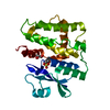
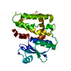
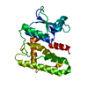
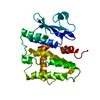
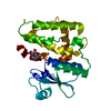
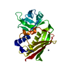
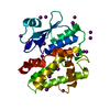
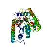
#1: Journal: J. Biol. Chem. / Year: 2002
Title: Functional divergence in the glutathione transferase superfamily in plants. Identification of two classes with putative functions in redox homeostasis in Arabidopsis thaliana.
Authors: Dixon, D.P. / Davis, B.G. / Edwards, R.
History
DepositionNov 4, 2015Deposition site: RCSB / Processing site: PDBE
Revision 1.0Nov 16, 2016Provider: repository / Type: Initial release

-
Structure visualization

Structure viewerMolecule:
MolmilJmol/JSmol

Downloads & links

-
Assembly

Deposited unit
A: Glutathione S-transferase DHAR1, mitochondrial
hetero molecules


Theoretical massNumber of molelcules
Total (without water)25,4967
Polymers24,7281
Non-polymers7686
Water4,378243
1


  • Idetical with deposited unit
  • defined by software
TypeNameSymmetry operationNumber
identity operation1_555x,y,z1
Buried area1640 Å2
ΔGint-8 kcal/mol
Surface area10680 Å2
MethodPISA
Unit cell
Length a, b, c (Å)63.920, 63.920, 266.260
Angle α, β, γ (deg.)90.00, 90.00, 120.00
Int Tables number179
Space group name H-MP6522
Components on special symmetry positions
IDModelComponents
11A-625-

HOH

-
Components

#1: Protein Glutathione S-transferase DHAR1, mitochondrial / Chloride intracellular channel homolog 1 / CLIC homolog 1 / Glutathione-dependent dehydroascorbate ...Chloride intracellular channel homolog 1 / CLIC homolog 1 / Glutathione-dependent dehydroascorbate reductase 1 / mtDHAR


Mass: 24728.123 Da / Num. of mol.: 1
Source method: isolated from a genetically manipulated source
Source: (gene. exp.) Arabidopsis thaliana (thale cress) / Gene: DHAR1, DHAR5, At1g19570, F14P1.9, F18O14.22 / Plasmid: pET24-DHAR1 / Production host: Escherichia coli BL21(DE3) (bacteria) / References: UniProt: Q9FWR4, glutathione transferase
#2: Chemical
ChemComp-GOL / GLYCEROL / GLYCERIN / PROPANE-1,2,3-TRIOL / Glycerol


Mass: 92.094 Da / Num. of mol.: 5 / Source method: obtained synthetically / Formula: C3H8O3
#3: Chemical ChemComp-GSH / GLUTATHIONE / Glutathione


Mass: 307.323 Da / Num. of mol.: 1 / Source method: obtained synthetically / Formula: C10H17N3O6S
#4: Water ChemComp-HOH / water / Water


Mass: 18.015 Da / Num. of mol.: 243 / Source method: isolated from a natural source / Formula: H2O

-
Experimental details

-
Experiment

ExperimentMethod: X-RAY DIFFRACTION

-
Sample preparation

CrystalDensity Matthews: 3.19 Å3/Da / Density % sol: 61.45 % / Description: hexagonal bipyramidal
Crystal growTemperature: 293 K / Method: vapor diffusion, sitting drop / pH: 7.5
Details: Protein 20 mg/ml 20% PEG 8000 0.2M NaK Phosphate 0.1M TRIS
PH range: 7.0 - 8.5

-
Data collection

DiffractionMean temperature: 100 K
Diffraction sourceSource: SYNCHROTRON / Site: Diamond / Beamline: I02 / Wavelength: 0.979 Å
DetectorType: DECTRIS PILATUS 6M-F / Detector: PIXEL / Date: Mar 10, 2014
RadiationMonochromator: double crystal Si(111) / Protocol: SINGLE WAVELENGTH / Monochromatic (M) / Laue (L): M / Scattering type: x-ray
Radiation wavelengthWavelength: 0.979 Å / Relative weight: 1
ReflectionResolution: 1.8→55.3 Å / Num. obs: 30540 / % possible obs: 99.7 % / Redundancy: 9.4 % / Net I/σ(I): 30
Reflection shellResolution: 1.81→1.86 Å / Redundancy: 9.8 % / Rmerge(I) obs: 0.8 / Mean I/σ(I) obs: 3.3 / % possible all: 99.3

-
Processing

Software
NameVersionClassification
REFMAC5.8.0073refinement
XDSdata reduction
Aimless0.2.12data scaling
RefinementResolution: 1.81→55.3 Å / Cor.coef. Fo:Fc: 0.971 / Cor.coef. Fo:Fc free: 0.96 / SU B: 3.924 / SU ML: 0.064 / SU R Cruickshank DPI: 0.0907 / Cross valid method: THROUGHOUT / ESU R: 0.091 / ESU R Free: 0.094 / SU Rfree Cruickshank DPI: 0.0939 / Stereochemistry target values: MAXIMUM LIKELIHOOD / Details: HYDROGENS HAVE BEEN ADDED IN THE RIDING POSITIONS
RfactorNum. reflection% reflectionSelection details
Rfree0.19538 1540 5.1 %RANDOM
Rwork0.16107 ---
obs0.16272 28950 99.57 %-
Solvent computationIon probe radii: 0.8 Å / Shrinkage radii: 0.8 Å / VDW probe radii: 1.2 Å / Solvent model: BABINET MODEL WITH MASK
Displacement parametersBiso mean: 44.177 Å2
Baniso -1Baniso -2Baniso -3
1-0.17 Å20.09 Å2-0 Å2
2--0.17 Å2-0 Å2
3----0.55 Å2
Refinement stepCycle: 1 / Resolution: 1.81→55.3 Å
ProteinNucleic acidLigandSolventTotal
Num. atoms1659 0 50 243 1952
Refine LS restraints
Refine-IDTypeDev idealDev ideal targetNumber
X-RAY DIFFRACTIONr_bond_refined_d0.0150.0191767
X-RAY DIFFRACTIONr_bond_other_d0.0010.021681
X-RAY DIFFRACTIONr_angle_refined_deg1.6241.9832393
X-RAY DIFFRACTIONr_angle_other_deg0.8633911
X-RAY DIFFRACTIONr_dihedral_angle_1_deg5.6735215
X-RAY DIFFRACTIONr_dihedral_angle_2_deg29.36725.35271
X-RAY DIFFRACTIONr_dihedral_angle_3_deg12.27215290
X-RAY DIFFRACTIONr_dihedral_angle_4_deg8.685152
X-RAY DIFFRACTIONr_chiral_restr0.110.2269
X-RAY DIFFRACTIONr_gen_planes_refined0.0080.0211928
X-RAY DIFFRACTIONr_gen_planes_other0.0010.02370
X-RAY DIFFRACTIONr_nbd_refined
X-RAY DIFFRACTIONr_nbd_other
X-RAY DIFFRACTIONr_nbtor_refined
X-RAY DIFFRACTIONr_nbtor_other
X-RAY DIFFRACTIONr_xyhbond_nbd_refined
X-RAY DIFFRACTIONr_xyhbond_nbd_other
X-RAY DIFFRACTIONr_metal_ion_refined
X-RAY DIFFRACTIONr_metal_ion_other
X-RAY DIFFRACTIONr_symmetry_vdw_refined
X-RAY DIFFRACTIONr_symmetry_vdw_other
X-RAY DIFFRACTIONr_symmetry_hbond_refined
X-RAY DIFFRACTIONr_symmetry_hbond_other
X-RAY DIFFRACTIONr_symmetry_metal_ion_refined
X-RAY DIFFRACTIONr_symmetry_metal_ion_other
X-RAY DIFFRACTIONr_mcbond_it1.9682.902854
X-RAY DIFFRACTIONr_mcbond_other1.9542.9853
X-RAY DIFFRACTIONr_mcangle_it2.5984.3371068
X-RAY DIFFRACTIONr_mcangle_other2.5974.3411069
X-RAY DIFFRACTIONr_scbond_it3.4993.41913
X-RAY DIFFRACTIONr_scbond_other3.4383.411913
X-RAY DIFFRACTIONr_scangle_it
X-RAY DIFFRACTIONr_scangle_other5.0474.9211325
X-RAY DIFFRACTIONr_long_range_B_refined8.51226.7942216
X-RAY DIFFRACTIONr_long_range_B_other8.49226.7962216
X-RAY DIFFRACTIONr_rigid_bond_restr
X-RAY DIFFRACTIONr_sphericity_free
X-RAY DIFFRACTIONr_sphericity_bonded
LS refinement shellResolution: 1.81→1.857 Å / Total num. of bins used: 20
RfactorNum. reflection% reflection
Rfree0.239 119 -
Rwork0.218 2063 -
obs--98.96 %
Refinement TLS params.Method: refined / Origin x: -28.0814 Å / Origin y: 16.9021 Å / Origin z: -8.1316 Å
111213212223313233
T0.0397 Å2-0.0201 Å20.0116 Å2-0.1142 Å20.0129 Å2--0.0256 Å2
L1.2587 °2-0.0943 °20.0571 °2-1.776 °20.9551 °2--1.314 °2
S-0.0102 Å °-0.0121 Å °-0.0094 Å °0.0626 Å °0.0534 Å °-0.121 Å °0.1465 Å °0.0209 Å °-0.0432 Å °

+
About Yorodumi

-
News

-
Feb 9, 2022. New format data for meta-information of EMDB entries

New format data for meta-information of EMDB entries

  • Version 3 of the EMDB header file is now the official format.
  • The previous official version 1.9 will be removed from the archive.

Related info.:EMDB header

External links:wwPDB to switch to version 3 of the EMDB data model

-
Aug 12, 2020. Covid-19 info

Covid-19 info

URL: https://pdbj.org/emnavi/covid19.php

New page: Covid-19 featured information page in EM Navigator.

Related info.:Covid-19 info / Mar 5, 2020. Novel coronavirus structure data

+
Mar 5, 2020. Novel coronavirus structure data

Novel coronavirus structure data

Related info.:Yorodumi Speices / Aug 12, 2020. Covid-19 info

External links:COVID-19 featured content - PDBj / Molecule of the Month (242):Coronavirus Proteases

+
Jan 31, 2019. EMDB accession codes are about to change! (news from PDBe EMDB page)

EMDB accession codes are about to change! (news from PDBe EMDB page)

  • The allocation of 4 digits for EMDB accession codes will soon come to an end. Whilst these codes will remain in use, new EMDB accession codes will include an additional digit and will expand incrementally as the available range of codes is exhausted. The current 4-digit format prefixed with “EMD-” (i.e. EMD-XXXX) will advance to a 5-digit format (i.e. EMD-XXXXX), and so on. It is currently estimated that the 4-digit codes will be depleted around Spring 2019, at which point the 5-digit format will come into force.
  • The EM Navigator/Yorodumi systems omit the EMD- prefix.

Related info.:Q: What is EMD? / ID/Accession-code notation in Yorodumi/EM Navigator

External links:EMDB Accession Codes are Changing Soon! / Contact to PDBj

+
Jul 12, 2017. Major update of PDB

Major update of PDB

  • wwPDB released updated PDB data conforming to the new PDBx/mmCIF dictionary.
  • This is a major update changing the version number from 4 to 5, and with Remediation, in which all the entries are updated.
  • In this update, many items about electron microscopy experimental information are reorganized (e.g. em_software).
  • Now, EM Navigator and Yorodumi are based on the updated data.

External links:wwPDB Remediation / Enriched Model Files Conforming to OneDep Data Standards Now Available in the PDB FTP Archive

-
Yorodumi

Thousand views of thousand structures

  • Yorodumi is a browser for structure data from EMDB, PDB, SASBDB, etc.
  • This page is also the successor to EM Navigator detail page, and also detail information page/front-end page for Omokage search.
  • The word "yorodu" (or yorozu) is an old Japanese word meaning "ten thousand". "mi" (miru) is to see.

Related info.:EMDB / PDB / SASBDB / Comparison of 3 databanks / Yorodumi Search / Aug 31, 2016. New EM Navigator & Yorodumi / Yorodumi Papers / Jmol/JSmol / Function and homology information / Changes in new EM Navigator and Yorodumi

Read more