 Movie
Movie Controller

[English] 日本語
 Yorodumi
- PDB-6fxi: Human PARP10 (ARTD10), catalytic fragment in complex with 3-amino... -
+ Open data

- Basic information
| Entry | Database: PDB / ID: 6fxi | ||||||
|---|---|---|---|---|---|---|---|
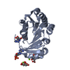
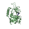
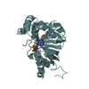
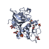
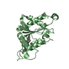
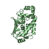
| Title | Human PARP10 (ARTD10), catalytic fragment in complex with 3-aminobenzamide and citrate | ||||||
 Components Components | Poly [ADP-ribose] polymerase 10 | ||||||
 Keywords Keywords |  TRANSFERASE / Transferase domain / ADP-ribosylation / PARP inhibitor TRANSFERASE / Transferase domain / ADP-ribosylation / PARP inhibitor | ||||||
| Function / homology |  Function and homology information Function and homology informationnegative regulation of protein K63-linked ubiquitination / NAD+- protein-lysine ADP-ribosyltransferase activity / NAD biosynthesis via nicotinamide riboside salvage pathway / Nicotinamide salvaging / Maturation of nucleoprotein / Maturation of nucleoprotein / K63-linked polyubiquitin modification-dependent protein binding / protein auto-ADP-ribosylation / protein poly-ADP-ribosylation / negative regulation of NF-kappaB transcription factor activity ...negative regulation of protein K63-linked ubiquitination / NAD+- protein-lysine ADP-ribosyltransferase activity / NAD biosynthesis via nicotinamide riboside salvage pathway / Nicotinamide salvaging / Maturation of nucleoprotein / Maturation of nucleoprotein / K63-linked polyubiquitin modification-dependent protein binding / protein auto-ADP-ribosylation / protein poly-ADP-ribosylation / negative regulation of NF-kappaB transcription factor activity / NAD+-protein ADP-ribosyltransferase activity / NAD+ ADP-ribosyltransferase activity / Transferases; Glycosyltransferases; Pentosyltransferases / translesion synthesis / negative regulation of fibroblast proliferation / nucleotidyltransferase activity / transcription corepressor activity / chromatin organization / DNA-binding transcription factor binding / viral protein processing / negative regulation of gene expression / nucleolus / Golgi apparatus / nucleus / cytosol / cytoplasmSimilarity search - Function | ||||||
| Biological species |  Homo sapiens (human) Homo sapiens (human) | ||||||
| Method | X-RAY DIFFRACTION / SYNCHROTRON / MOLECULAR REPLACEMENT / Resolution: 2.6 Å | ||||||
 Authors Authors | Karlberg, T. / Thorsell, A.G. / Schuler, H. | ||||||
 Citation Citation | Journal: To Be Published Title: Human PARP10 (ARTD10), catalytic fragment in complex with 3-aminobenzamide and citrate Authors: Karlberg, T. / Thorsell, A.G. / Schuler, H. | ||||||
| History |
|
- Structure visualization
| Structure viewer | Molecule: MolmilJmol/JSmol |
|---|
- Downloads & links
-Download
| PDBx/mmCIF format | 6fxi.cif.gz | 166.1 KB | Display | PDBx/mmCIF format |
|---|---|---|---|---|
| PDB format | pdb6fxi.ent.gz | 131.9 KB | Display | PDB format |
| PDBx/mmJSON format | 6fxi.json.gz | Tree view | PDBx/mmJSON format | |
| Others |  Other downloads Other downloads |
-Validation report
| Arichive directory | https://data.pdbj.org/pub/pdb/validation_reports/fx/6fxiftp://data.pdbj.org/pub/pdb/validation_reports/fx/6fxi | HTTPS FTP |
|---|
-Related structure data
| Related structure data | 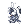 5lx6S S: Starting model for refinement |
|---|---|
| Similar structure data |


-Links
 PDBj
PDBj- Assembly
| Deposited unit | 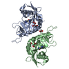
| ||||||||
|---|---|---|---|---|---|---|---|---|---|
| 1 | 
| ||||||||
| 2 | 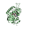
| ||||||||
| Unit cell |
|
-Components
| #1: Protein | Mass: 21628.586 Da / Num. of mol.: 2 Source method: isolated from a genetically manipulated source Source: (gene. exp.) Homo sapiens (human) / Gene: PARP10 / Plasmid: pNIC-Bsa28 / Production host:  Escherichia coli (E. coli) / References: UniProt: Q53GL7, NAD+ ADP-ribosyltransferase Escherichia coli (E. coli) / References: UniProt: Q53GL7, NAD+ ADP-ribosyltransferase#2: Chemical | 3-Aminobenzamide  Mass: 136.151 Da / Num. of mol.: 2 / Source method: obtained synthetically / Formula: C7H8N2O Mass: 136.151 Da / Num. of mol.: 2 / Source method: obtained synthetically / Formula: C7H8N2O#3: Chemical | Citric acid  Mass: 192.124 Da / Num. of mol.: 2 / Source method: obtained synthetically / Formula: C6H8O7 Mass: 192.124 Da / Num. of mol.: 2 / Source method: obtained synthetically / Formula: C6H8O7#4: Chemical | ChemComp-GOL / | Glycerol  Mass: 92.094 Da / Num. of mol.: 1 / Source method: obtained synthetically / Formula: C3H8O3 Mass: 92.094 Da / Num. of mol.: 1 / Source method: obtained synthetically / Formula: C3H8O3#5: Water | ChemComp-HOH / | Water Mass: 18.015 Da / Num. of mol.: 45 / Source method: isolated from a natural source / Formula: H2O Mass: 18.015 Da / Num. of mol.: 45 / Source method: isolated from a natural source / Formula: H2O |
|---|
-Experimental details
-Experiment
| Experiment | Method: X-RAY DIFFRACTION / Number of used crystals: 1 |
|---|
- Sample preparation
| Crystal | Density Matthews: 2.87 Å3/Da / Density % sol: 57.1 % |
|---|---|
| Crystal grow | Temperature: 277 K / Method: vapor diffusion, sitting drop / pH: 5 Details: 16% PEG3350, 0.1 M Citrate-Bis-Tris-Propane, 3 mM 3-aminobenzamide |
-Data collection
| Diffraction | Mean temperature: 100 K |
|---|---|
| Diffraction source | Source: SYNCHROTRON / Site: Diamond  / Beamline: I04 / Wavelength: 0.9795 Å / Beamline: I04 / Wavelength: 0.9795 Å |
| Detector | Type: DECTRIS PILATUS 6M-F / Detector: PIXEL / Date: Sep 21, 2015 |
| Radiation | Protocol: SINGLE WAVELENGTH / Monochromatic (M) / Laue (L): M / Scattering type: x-ray |
| Radiation wavelength | Wavelength: 0.9795 Å / Relative weight: 1 |
| Reflection | Resolution: 2.6→49.85 Å / Num. obs: 15114 / % possible obs: 98.8 % / Observed criterion σ(F): 0 / Observed criterion σ(I): 0 / Redundancy: 6.9 % / Biso Wilson estimate: 50.17 Å2 / CC1/2: 0.985 / Rmerge(I) obs: 0.223 / Rrim(I) all: 0.242 / Net I/σ(I): 7.9 |
| Reflection shell | Resolution: 2.6→2.71 Å / Redundancy: 6.9 % / Rmerge(I) obs: 0.737 / Num. unique obs: 1747 / CC1/2: 0.864 / Rrim(I) all: 0.797 / % possible all: 100 |
- Processing
| Software |
| ||||||||||||||||||||||||||||||||||||||||||||||||||||||||||||||||||||||||||||||||||||||||||||||||||||||||||||||||||
|---|---|---|---|---|---|---|---|---|---|---|---|---|---|---|---|---|---|---|---|---|---|---|---|---|---|---|---|---|---|---|---|---|---|---|---|---|---|---|---|---|---|---|---|---|---|---|---|---|---|---|---|---|---|---|---|---|---|---|---|---|---|---|---|---|---|---|---|---|---|---|---|---|---|---|---|---|---|---|---|---|---|---|---|---|---|---|---|---|---|---|---|---|---|---|---|---|---|---|---|---|---|---|---|---|---|---|---|---|---|---|---|---|---|---|---|
| Refinement | Method to determine structure: MOLECULAR REPLACEMENT Starting model: 5LX6 Resolution: 2.6→49.85 Å / Cor.coef. Fo:Fc: 0.887 / Cor.coef. Fo:Fc free: 0.853 / SU R Cruickshank DPI: 0.593 / Cross valid method: THROUGHOUT / σ(F): 0 / SU R Blow DPI: 0.541 / SU Rfree Blow DPI: 0.28 / SU Rfree Cruickshank DPI: 0.289
| ||||||||||||||||||||||||||||||||||||||||||||||||||||||||||||||||||||||||||||||||||||||||||||||||||||||||||||||||||
| Displacement parameters | Biso mean: 36.8 Å2
| ||||||||||||||||||||||||||||||||||||||||||||||||||||||||||||||||||||||||||||||||||||||||||||||||||||||||||||||||||
| Refine analyze | Luzzati coordinate error obs: 0.36 Å | ||||||||||||||||||||||||||||||||||||||||||||||||||||||||||||||||||||||||||||||||||||||||||||||||||||||||||||||||||
| Refinement step | Cycle: 1 / Resolution: 2.6→49.85 Å
| ||||||||||||||||||||||||||||||||||||||||||||||||||||||||||||||||||||||||||||||||||||||||||||||||||||||||||||||||||
| Refine LS restraints |
| ||||||||||||||||||||||||||||||||||||||||||||||||||||||||||||||||||||||||||||||||||||||||||||||||||||||||||||||||||
| LS refinement shell | Resolution: 2.6→2.78 Å / Total num. of bins used: 8
| ||||||||||||||||||||||||||||||||||||||||||||||||||||||||||||||||||||||||||||||||||||||||||||||||||||||||||||||||||
| Refinement TLS params. | Method: refined / Refine-ID: X-RAY DIFFRACTION
| ||||||||||||||||||||||||||||||||||||||||||||||||||||||||||||||||||||||||||||||||||||||||||||||||||||||||||||||||||
| Refinement TLS group |
|
