[English] 日本語
Yorodumi
- PDB-5una: Fragment of 7SK snRNA methylphosphate capping enzyme -

+
Open data


ID or keywords:

Loading...

-
Basic information

Entry
Database: PDB / ID: 5una
TitleFragment of 7SK snRNA methylphosphate capping enzyme
Components
  • 7SK snRNA methylphosphate capping enzyme
  • unidentified peptide section/fragment
KeywordsTRANSFERASE / Structural Genomics Consortium (SGC) / methyltransferase
Function / homology
Function and homology information


RNA 5'-gamma-phosphate methyltransferase activity / snRNA metabolic process / snRNA modification / 7SK snRNP / positive regulation of snRNA transcription by RNA polymerase II / 7SK snRNA binding / snRNA binding / positive regulation of protein localization to Cajal body / RNA methyltransferase activity / RNA methylation ...RNA 5'-gamma-phosphate methyltransferase activity / snRNA metabolic process / snRNA modification / 7SK snRNP / positive regulation of snRNA transcription by RNA polymerase II / 7SK snRNA binding / snRNA binding / positive regulation of protein localization to Cajal body / RNA methyltransferase activity / RNA methylation / O-methyltransferase activity / S-adenosylmethionine-dependent methyltransferase activity / positive regulation of G1/S transition of mitotic cell cycle / Transferases; Transferring one-carbon groups; Methyltransferases / ribonucleoprotein complex / negative regulation of transcription by RNA polymerase II / RNA binding / nucleus
Similarity search - Function
RNA methyltransferase bin3, C-terminal / Bin3-type S-adenosyl-L-methionine binding domain / RNA methyltransferase Bin3-like / Bicoid-interacting protein 3 (Bin3) / Bin3-type S-adenosyl-L-methionine (SAM) domain profile. / Methyltransferase domain / Vaccinia Virus protein VP39 / S-adenosyl-L-methionine-dependent methyltransferase superfamily / Rossmann fold / 3-Layer(aba) Sandwich / Alpha Beta
Similarity search - Domain/homology
S-ADENOSYL-L-HOMOCYSTEINE / 7SK snRNA methylphosphate capping enzyme
Similarity search - Component
Biological speciesHomo sapiens (human)
MethodX-RAY DIFFRACTION / SYNCHROTRON / MAD / Resolution: 2.55 Å
AuthorsWu, H. / Tempel, W. / Dombrovski, L. / McCarthy, A.A. / Loppnau, P. / Bountra, C. / Arrowsmith, C.H. / Edwards, A.M. / Park, H. / Structural Genomics Consortium (SGC)
CitationJournal: To Be Published
Title: Fragment of 7SK snRNA methylphosphate capping enzyme
Authors: Wu, H. / Tempel, W. / Dombrovski, L. / McCarthy, A.A. / Loppnau, P. / Bountra, C. / Arrowsmith, C.H. / Edwards, A.M. / Park, H. / Structural Genomics Consortium (SGC)
History
DepositionJan 30, 2017Deposition site: RCSB / Processing site: RCSB
SupersessionMar 8, 2017ID: 3G07
Revision 1.0Mar 8, 2017Provider: repository / Type: Initial release

-
Structure visualization

Structure viewerMolecule:
MolmilJmol/JSmol

Downloads & links

-
Assembly

Deposited unit
A: 7SK snRNA methylphosphate capping enzyme
B: 7SK snRNA methylphosphate capping enzyme
C: 7SK snRNA methylphosphate capping enzyme
D: 7SK snRNA methylphosphate capping enzyme
E: 7SK snRNA methylphosphate capping enzyme
F: 7SK snRNA methylphosphate capping enzyme
M: unidentified peptide section/fragment
N: unidentified peptide section/fragment
O: unidentified peptide section/fragment
Q: unidentified peptide section/fragment
R: unidentified peptide section/fragment
hetero molecules


Theoretical massNumber of molelcules
Total (without water)204,91517
Polymers202,60811
Non-polymers2,3066
Water0
1
A: 7SK snRNA methylphosphate capping enzyme
hetero molecules


Theoretical massNumber of molelcules
Total (without water)33,7122
Polymers33,3281
Non-polymers3841
Water0
TypeNameSymmetry operationNumber
identity operation1_555x,y,z1
MethodPISA
2
B: 7SK snRNA methylphosphate capping enzyme
hetero molecules


Theoretical massNumber of molelcules
Total (without water)33,7122
Polymers33,3281
Non-polymers3841
Water0
TypeNameSymmetry operationNumber
identity operation1_555x,y,z1
MethodPISA
3
C: 7SK snRNA methylphosphate capping enzyme
hetero molecules


Theoretical massNumber of molelcules
Total (without water)33,7122
Polymers33,3281
Non-polymers3841
Water0
TypeNameSymmetry operationNumber
identity operation1_555x,y,z1
MethodPISA
4
D: 7SK snRNA methylphosphate capping enzyme
hetero molecules


Theoretical massNumber of molelcules
Total (without water)33,7122
Polymers33,3281
Non-polymers3841
Water0
TypeNameSymmetry operationNumber
identity operation1_555x,y,z1
MethodPISA
5
E: 7SK snRNA methylphosphate capping enzyme
hetero molecules


Theoretical massNumber of molelcules
Total (without water)33,7122
Polymers33,3281
Non-polymers3841
Water0
TypeNameSymmetry operationNumber
identity operation1_555x,y,z1
MethodPISA
6
F: 7SK snRNA methylphosphate capping enzyme
hetero molecules


Theoretical massNumber of molelcules
Total (without water)33,7122
Polymers33,3281
Non-polymers3841
Water0
TypeNameSymmetry operationNumber
identity operation1_555x,y,z1
MethodPISA
7
M: unidentified peptide section/fragment


  • defined by software
  • 529 Da, 1 polymers
Theoretical massNumber of molelcules
Total (without water)5291
Polymers5291
Non-polymers00
Water0
TypeNameSymmetry operationNumber
identity operation1_555x,y,z1
MethodPISA
8
N: unidentified peptide section/fragment


  • defined by software
  • 529 Da, 1 polymers
Theoretical massNumber of molelcules
Total (without water)5291
Polymers5291
Non-polymers00
Water0
TypeNameSymmetry operationNumber
identity operation1_555x,y,z1
MethodPISA
9
O: unidentified peptide section/fragment


  • defined by software
  • 529 Da, 1 polymers
Theoretical massNumber of molelcules
Total (without water)5291
Polymers5291
Non-polymers00
Water0
TypeNameSymmetry operationNumber
identity operation1_555x,y,z1
MethodPISA
10
Q: unidentified peptide section/fragment


  • defined by software
  • 529 Da, 1 polymers
Theoretical massNumber of molelcules
Total (without water)5291
Polymers5291
Non-polymers00
Water0
TypeNameSymmetry operationNumber
identity operation1_555x,y,z1
MethodPISA
11
R: unidentified peptide section/fragment


  • defined by software
  • 529 Da, 1 polymers
Theoretical massNumber of molelcules
Total (without water)5291
Polymers5291
Non-polymers00
Water0
TypeNameSymmetry operationNumber
identity operation1_555x,y,z1
MethodPISA
Unit cell
Length a, b, c (Å)183.240, 183.240, 151.390
Angle α, β, γ (deg.)90.000, 90.000, 120.000
Int Tables number146
Space group name H-MH3

-
Components

#1: Protein
7SK snRNA methylphosphate capping enzyme / MePCE / Bicoid-interacting protein 3 homolog / Bin3 homolog


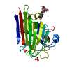
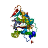
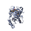
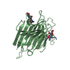
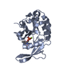
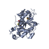
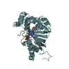
Mass: 33327.508 Da / Num. of mol.: 6
Source method: isolated from a genetically manipulated source
Source: (gene. exp.) Homo sapiens (human) / Gene: MEPCE, BCDIN3 / Plasmid: pET28a-LIC / Production host: Escherichia coli (E. coli) / Strain (production host): BL21 (DE3) codon plus RIL
References: UniProt: Q7L2J0, Transferases; Transferring one-carbon groups; Methyltransferases
#2: Protein/peptide
unidentified peptide section/fragment


Mass: 528.644 Da / Num. of mol.: 5
Source method: isolated from a genetically manipulated source
Source: (gene. exp.) Homo sapiens (human) / Production host: Escherichia coli (E. coli)
#3: Chemical
ChemComp-SAH / S-ADENOSYL-L-HOMOCYSTEINE / S-Adenosyl-L-homocysteine


Type: L-peptide linking / Mass: 384.411 Da / Num. of mol.: 6 / Source method: obtained synthetically / Formula: C14H20N6O5S

-
Experimental details

-
Experiment

ExperimentMethod: X-RAY DIFFRACTION / Number of used crystals: 2

-
Sample preparation

Crystal
IDDensity Matthews3/Da)Density % sol (%)
12.4149.05
2
Crystal grow
Temperature (K)Crystal-IDMethodpHDetails
2931vapor diffusion, hanging drop8Pre-incubated with SAM in 10-fold stoichiometric excess. Reservoir: 22% PEG 3350, 0.1 M Ammonium Sulfate, 0.1 M Tris-HCl.
2912vapor diffusion, hanging drop8Purified protein (10 mg/mL) was complexed with S-adenosyl-L-methionine in a 1:10 stoichiometric ratio. This solution (2 muL) was then mixed with precipitant: 22% PEG 3350, 0.1 M ammonium sulfate, 0.1 M TRIS-HCl.

-
Data collection

DiffractionMean temperature: 100 K
Diffraction sourceSource: SYNCHROTRON / Site: ESRF / Beamline: ID14-4 / Wavelength: 0.976 Å
DetectorType: ADSC QUANTUM 315 / Detector: CCD / Date: Sep 22, 2008
RadiationProtocol: MAD / Monochromatic (M) / Laue (L): M / Scattering type: x-ray
Radiation wavelengthWavelength: 0.976 Å / Relative weight: 1
ReflectionResolution: 2.5→44.2 Å / Num. obs: 65295 / % possible obs: 99.6 % / Redundancy: 5.7 % / Biso Wilson estimate: 81.56 Å2 / CC1/2: 0.999 / Rmerge(I) obs: 0.062 / Rpim(I) all: 0.029 / Rrim(I) all: 0.068 / Net I/σ(I): 18.3 / Num. measured all: 375144 / Scaling rejects: 0
Reflection shell

Diffraction-ID: 1

Resolution (Å)Redundancy (%)Rmerge(I) obsNum. measured allNum. unique allCC1/2Rpim(I) allRrim(I) allNet I/σ(I) obs% possible all
2.5-2.565.81.1012682646450.6160.5041.2121.6100
11.46-44.24.60.04722654960.9940.0250.05445.173.5

-
Processing

Software
NameVersionClassification
BUSTER2.10.2refinement
Aimless0.5.28data scaling
PDB_EXTRACT3.22data extraction
XDSdata reduction
SOLVEphasing
RefinementMethod to determine structure: MAD / Resolution: 2.55→25.7 Å / Cor.coef. Fo:Fc: 0.943 / Cor.coef. Fo:Fc free: 0.937 / Rfactor Rfree error: 0 / SU R Cruickshank DPI: 0.337 / Cross valid method: THROUGHOUT / σ(F): 0 / SU R Blow DPI: 0.3 / SU Rfree Blow DPI: 0.207 / SU Rfree Cruickshank DPI: 0.219
Details: this model supersedes PDB entry 3g07. Compared to the superseded entry, diffraction data have ben re-processed and free reflections have been reassigned. Due to dicontinuities in electron ...Details: this model supersedes PDB entry 3g07. Compared to the superseded entry, diffraction data have ben re-processed and free reflections have been reassigned. Due to dicontinuities in electron density, N-terminal segments of the model may have been assigned to incorrect protein chains. Weak electron density suggests Cys463 may form a disulfide bridge to another cystyl residue, possibly Cys522. Overall, poorly contoured density in several areas of the map may have lead to incorrect register with respect to the amino acid sequence. Non crystallographic symmetry was applied during map interpretation and restrained coordinate refinement.Some residues not or poorly supported by electron density, such as A684 in chain C and Y637 in chain A, were included in the model when their probable positions could be inferred from non crystallographic symmetry or density supporting respective up and downstream residues.
RfactorNum. reflection% reflectionSelection details
Rfree0.217 1909 3.1 %CCP4 FREERFLAG
Rwork0.204 ---
obs0.204 61497 99.6 %-
Displacement parametersBiso max: 168.52 Å2 / Biso mean: 90.63 Å2 / Biso min: 37.49 Å2
Baniso -1Baniso -2Baniso -3
1-2.6238 Å20 Å20 Å2
2--2.6238 Å20 Å2
3----5.2476 Å2
Refine analyzeLuzzati coordinate error obs: 0.35 Å
Refinement stepCycle: final / Resolution: 2.55→25.7 Å
ProteinNucleic acidLigandSolventTotal
Num. atoms9476 0 179 0 9655
Biso mean--79.32 --
Num. residues----1271
Refine LS restraints
Refine-IDTypeNumberRestraint functionWeightDev ideal
X-RAY DIFFRACTIONt_dihedral_angle_d3086SINUSOIDAL2
X-RAY DIFFRACTIONt_trig_c_planes168HARMONIC2
X-RAY DIFFRACTIONt_gen_planes1573HARMONIC5
X-RAY DIFFRACTIONt_it9883HARMONIC20
X-RAY DIFFRACTIONt_nbd
X-RAY DIFFRACTIONt_improper_torsion
X-RAY DIFFRACTIONt_pseud_angle
X-RAY DIFFRACTIONt_chiral_improper_torsion1275SEMIHARMONIC5
X-RAY DIFFRACTIONt_sum_occupancies
X-RAY DIFFRACTIONt_utility_distance
X-RAY DIFFRACTIONt_utility_angle
X-RAY DIFFRACTIONt_utility_torsion
X-RAY DIFFRACTIONt_ideal_dist_contact10665SEMIHARMONIC4
X-RAY DIFFRACTIONt_bond_d9883HARMONIC20.01
X-RAY DIFFRACTIONt_angle_deg13503HARMONIC21.07
X-RAY DIFFRACTIONt_omega_torsion3.02
X-RAY DIFFRACTIONt_other_torsion18.86
LS refinement shellResolution: 2.55→2.62 Å / Total num. of bins used: 20
RfactorNum. reflection% reflection
Rfree0.259 124 2.71 %
Rwork0.244 4450 -
all-4574 -
obs--100 %
Refinement TLS params.

Method: refined / Refine-ID: X-RAY DIFFRACTION

IDL112)L122)L132)L222)L232)L332)S11 (Å °)S12 (Å °)S13 (Å °)S21 (Å °)S22 (Å °)S23 (Å °)S31 (Å °)S32 (Å °)S33 (Å °)T112)T122)T132)T222)T232)T332)Origin x (Å)Origin y (Å)Origin z (Å)
14.28740.56471.17124.40491.43443.0001-0.0183-0.26410.61231.3689-0.2817-0.64290.35340.15030.29990.0933-0.3501-0.252-0.067-0.0468-0.16837.182673.660958.194
23.16510.38982.39933.5328-0.0686.1030.5835-0.0664-0.2099-0.46010.10560.73621.04130.2348-0.6892-0.1747-0.0414-0.3344-0.2159-0.0287-0.03459.315471.628329.0112
35.23842.08862.11883.62080.45023.20170.32220.11310.07710.128-0.1663-0.27830.03531.2773-0.156-0.3095-0.2257-0.05380.6531-0.2335-0.2664-4.831933.1321-4.4447
44.173-0.2534-0.64871.5123-0.27423.56440.05280.93470.3923-0.02280.37730.20450.3497-0.9671-0.4301-0.3098-0.2245-0.15610.36230.2040.1659-21.827765.00531.9409
53.61840.4781-0.852.70841.33282.43040.13780.2334-0.10190.6229-0.51640.7150.5411-0.51990.3786-0.086-0.34330.10220.1417-0.292-0.155310.766155.75242.9852
63.3344-0.1134-0.6271.90561.23433.13470.0724-0.48861.48690.14460.5999-0.3984-0.24440.5766-0.6724-0.516-0.0632-0.07970.0267-0.3970.60796.873880.384815.5592
Refinement TLS group
IDRefine-IDRefine TLS-IDSelection detailsAuth asym-IDAuth seq-ID
1X-RAY DIFFRACTION1{ A|* }A413 - 685
2X-RAY DIFFRACTION2{ B|* }B413 - 686
3X-RAY DIFFRACTION3{ C|* }C413 - 686
4X-RAY DIFFRACTION4{ D|* }D412 - 686
5X-RAY DIFFRACTION5{ E|* }E413 - 686
6X-RAY DIFFRACTION6{ F|* }F412 - 686

+
About Yorodumi

-
News

-
Feb 9, 2022. New format data for meta-information of EMDB entries

New format data for meta-information of EMDB entries

  • Version 3 of the EMDB header file is now the official format.
  • The previous official version 1.9 will be removed from the archive.

Related info.:EMDB header

External links:wwPDB to switch to version 3 of the EMDB data model

-
Aug 12, 2020. Covid-19 info

Covid-19 info

URL: https://pdbj.org/emnavi/covid19.php

New page: Covid-19 featured information page in EM Navigator.

Related info.:Covid-19 info / Mar 5, 2020. Novel coronavirus structure data

+
Mar 5, 2020. Novel coronavirus structure data

Novel coronavirus structure data

Related info.:Yorodumi Speices / Aug 12, 2020. Covid-19 info

External links:COVID-19 featured content - PDBj / Molecule of the Month (242):Coronavirus Proteases

+
Jan 31, 2019. EMDB accession codes are about to change! (news from PDBe EMDB page)

EMDB accession codes are about to change! (news from PDBe EMDB page)

  • The allocation of 4 digits for EMDB accession codes will soon come to an end. Whilst these codes will remain in use, new EMDB accession codes will include an additional digit and will expand incrementally as the available range of codes is exhausted. The current 4-digit format prefixed with “EMD-” (i.e. EMD-XXXX) will advance to a 5-digit format (i.e. EMD-XXXXX), and so on. It is currently estimated that the 4-digit codes will be depleted around Spring 2019, at which point the 5-digit format will come into force.
  • The EM Navigator/Yorodumi systems omit the EMD- prefix.

Related info.:Q: What is EMD? / ID/Accession-code notation in Yorodumi/EM Navigator

External links:EMDB Accession Codes are Changing Soon! / Contact to PDBj

+
Jul 12, 2017. Major update of PDB

Major update of PDB

  • wwPDB released updated PDB data conforming to the new PDBx/mmCIF dictionary.
  • This is a major update changing the version number from 4 to 5, and with Remediation, in which all the entries are updated.
  • In this update, many items about electron microscopy experimental information are reorganized (e.g. em_software).
  • Now, EM Navigator and Yorodumi are based on the updated data.

External links:wwPDB Remediation / Enriched Model Files Conforming to OneDep Data Standards Now Available in the PDB FTP Archive

-
Yorodumi

Thousand views of thousand structures

  • Yorodumi is a browser for structure data from EMDB, PDB, SASBDB, etc.
  • This page is also the successor to EM Navigator detail page, and also detail information page/front-end page for Omokage search.
  • The word "yorodu" (or yorozu) is an old Japanese word meaning "ten thousand". "mi" (miru) is to see.

Related info.:EMDB / PDB / SASBDB / Comparison of 3 databanks / Yorodumi Search / Aug 31, 2016. New EM Navigator & Yorodumi / Yorodumi Papers / Jmol/JSmol / Function and homology information / Changes in new EM Navigator and Yorodumi

Read more