 Movie
Movie Controller

[English] 日本語
 Yorodumi
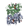
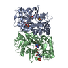
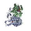
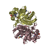
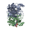
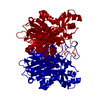
- PDB-6fko: Deoxyguanylosuccinate synthase (DgsS) quaternary structure with A... -
+ Open data

- Basic information
| Entry | Database: PDB / ID: 6fko | |||||||||
|---|---|---|---|---|---|---|---|---|---|---|
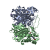
| Title | Deoxyguanylosuccinate synthase (DgsS) quaternary structure with ATP, dGMP, hadacidin at 2.1 Angstrom resolution | |||||||||
 Components Components | Adenylosuccinate synthetase | |||||||||
 Keywords Keywords | BIOSYNTHETIC PROTEIN / 2 / 6-diaminopurine / phage phiVC8 / Synthetase | |||||||||
| Function / homology |  Function and homology information Function and homology information2-amino-2'-deoxyadenylo-succinate synthase / adenylosuccinate synthase activity / IMP metabolic process / 'de novo' AMP biosynthetic process / purine nucleotide biosynthetic process / magnesium ion binding / ATP binding Similarity search - Function | |||||||||
| Biological species |  Vibrio phage phiVC8 (virus) Vibrio phage phiVC8 (virus) | |||||||||
| Method |  X-RAY DIFFRACTION / SYNCHROTRON / MOLECULAR REPLACEMENT / Resolution: 2.1 Å X-RAY DIFFRACTION / SYNCHROTRON / MOLECULAR REPLACEMENT / Resolution: 2.1 Å | |||||||||
 Authors Authors | Sleiman, D. / Loc'h, J. / Haouz, A. / Kaminski, P.A. | |||||||||
 Citation Citation | Journal: To Be Published Title: Deoxyguanylosuccinate synthase (DgsS) quaternary structure with ATP, dGMP, HAdacidin at 2.1 Angstrom resolution Authors: Sleiman, D. / Loc'h, J. / Haouz, A. / Kaminski, P.A. | |||||||||
| History |
|
- Structure visualization
| Structure viewer | Molecule: MolmilJmol/JSmol |
|---|
- Downloads & links
-Download
| PDBx/mmCIF format | 6fko.cif.gz | 288.1 KB | Display | PDBx/mmCIF format |
|---|---|---|---|---|
| PDB format | pdb6fko.ent.gz | 230.9 KB | Display | PDB format |
| PDBx/mmJSON format | 6fko.json.gz | Tree view | PDBx/mmJSON format | |
| Others |  Other downloads Other downloads |
-Validation report
| Arichive directory | https://data.pdbj.org/pub/pdb/validation_reports/fk/6fkoftp://data.pdbj.org/pub/pdb/validation_reports/fk/6fko | HTTPS FTP |
|---|
-Related structure data
| Similar structure data |
|---|
-Links
 PDBj
PDBj


- Assembly
| Deposited unit | 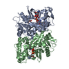
| ||||||||
|---|---|---|---|---|---|---|---|---|---|
| 1 |
| ||||||||
| Unit cell |
|
-Components
-Protein , 1 types, 2 molecules AB
| #1: Protein | Mass: 40392.996 Da / Num. of mol.: 2 Source method: isolated from a genetically manipulated source Source: (gene. exp.) Vibrio phage phiVC8 (virus) / Gene: phiVC8_p27 / Production host:  |
|---|
-Non-polymers , 5 types, 538 molecules 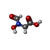


| #2: Chemical | ChemComp-HDA /  Mass: 119.076 Da / Num. of mol.: 1 / Source method: obtained synthetically / Formula: C3H5NO4 / Feature type: SUBJECT OF INVESTIGATION / Comment: anticancer, medication*YM Mass: 119.076 Da / Num. of mol.: 1 / Source method: obtained synthetically / Formula: C3H5NO4 / Feature type: SUBJECT OF INVESTIGATION / Comment: anticancer, medication*YM | ||||||
|---|---|---|---|---|---|---|---|
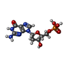
| #3: Chemical |  Mass: 347.221 Da / Num. of mol.: 2 / Source method: obtained synthetically / Formula: C10H14N5O7P / Feature type: SUBJECT OF INVESTIGATION Mass: 347.221 Da / Num. of mol.: 2 / Source method: obtained synthetically / Formula: C10H14N5O7P / Feature type: SUBJECT OF INVESTIGATION#4: Chemical | ChemComp-FLC / |  Mass: 189.100 Da / Num. of mol.: 1 / Source method: obtained synthetically / Formula: C6H5O7 Mass: 189.100 Da / Num. of mol.: 1 / Source method: obtained synthetically / Formula: C6H5O7#5: Chemical |  Mass: 507.181 Da / Num. of mol.: 2 / Source method: obtained synthetically / Formula: C10H16N5O13P3 / Feature type: SUBJECT OF INVESTIGATION / Comment: ATP, energy-carrying molecule*YM Mass: 507.181 Da / Num. of mol.: 2 / Source method: obtained synthetically / Formula: C10H16N5O13P3 / Feature type: SUBJECT OF INVESTIGATION / Comment: ATP, energy-carrying molecule*YM#6: Water | ChemComp-HOH / | Mass: 18.015 Da / Num. of mol.: 532 / Source method: isolated from a natural source / Formula: H2O |
-Experimental details
-Experiment
| Experiment | Method: X-RAY DIFFRACTION / Number of used crystals: 1 |
|---|
- Sample preparation
| Crystal | Density Matthews: 2.52 Å3/Da / Density % sol: 51.24 % |
|---|---|
| Crystal grow | Temperature: 291 K / Method: vapor diffusion / pH: 7.5 Details: 0.2 M (NH4)2SO4 0.1 M Na3 Cit 5.6 pH 25 %w/v PEG 4K |
-Data collection
| Diffraction | Mean temperature: 100 K |
|---|---|
| Diffraction source | Source: SYNCHROTRON / Site: SOLEIL  / Beamline: PROXIMA 2 / Wavelength: 0.980097 Å / Beamline: PROXIMA 2 / Wavelength: 0.980097 Å |
| Detector | Type: DECTRIS EIGER X 9M / Detector: PIXEL / Date: Jun 9, 2017 |
| Radiation | Protocol: SINGLE WAVELENGTH / Monochromatic (M) / Laue (L): M / Scattering type: x-ray |
| Radiation wavelength | Wavelength: 0.980097 Å / Relative weight: 1 |
| Reflection | Resolution: 2.106→48.188 Å / Num. obs: 49180 / % possible obs: 99.5 % / Redundancy: 5.91 % / Biso Wilson estimate: 48.18 Å2 / Net I/σ(I): 8.4 |
| Reflection shell | Resolution: 2.106→2.22 Å |
- Processing
| Software |
| ||||||||||||||||||||||||||||||||||||||||||||||||||||||||||||||||||||||||||||||||||||||||||||||||||||||||||||||||||
|---|---|---|---|---|---|---|---|---|---|---|---|---|---|---|---|---|---|---|---|---|---|---|---|---|---|---|---|---|---|---|---|---|---|---|---|---|---|---|---|---|---|---|---|---|---|---|---|---|---|---|---|---|---|---|---|---|---|---|---|---|---|---|---|---|---|---|---|---|---|---|---|---|---|---|---|---|---|---|---|---|---|---|---|---|---|---|---|---|---|---|---|---|---|---|---|---|---|---|---|---|---|---|---|---|---|---|---|---|---|---|---|---|---|---|---|
| Refinement | Method to determine structure: MOLECULAR REPLACEMENT / Resolution: 2.1→45.71 Å / Cor.coef. Fo:Fc: 0.948 / Cor.coef. Fo:Fc free: 0.931 / SU R Cruickshank DPI: 0.191 / Cross valid method: THROUGHOUT / σ(F): 0 / SU R Blow DPI: 0.212 / SU Rfree Blow DPI: 0.168 / SU Rfree Cruickshank DPI: 0.161
| ||||||||||||||||||||||||||||||||||||||||||||||||||||||||||||||||||||||||||||||||||||||||||||||||||||||||||||||||||
| Displacement parameters | Biso mean: 44.48 Å2
| ||||||||||||||||||||||||||||||||||||||||||||||||||||||||||||||||||||||||||||||||||||||||||||||||||||||||||||||||||
| Refine analyze | Luzzati coordinate error obs: 0.27 Å | ||||||||||||||||||||||||||||||||||||||||||||||||||||||||||||||||||||||||||||||||||||||||||||||||||||||||||||||||||
| Refinement step | Cycle: 1 / Resolution: 2.1→45.71 Å
| ||||||||||||||||||||||||||||||||||||||||||||||||||||||||||||||||||||||||||||||||||||||||||||||||||||||||||||||||||
| Refine LS restraints |
| ||||||||||||||||||||||||||||||||||||||||||||||||||||||||||||||||||||||||||||||||||||||||||||||||||||||||||||||||||
| LS refinement shell | Resolution: 2.1→2.15 Å / Total num. of bins used: 20
| ||||||||||||||||||||||||||||||||||||||||||||||||||||||||||||||||||||||||||||||||||||||||||||||||||||||||||||||||||
| Refinement TLS params. | Method: refined / Refine-ID: X-RAY DIFFRACTION
| ||||||||||||||||||||||||||||||||||||||||||||||||||||||||||||||||||||||||||||||||||||||||||||||||||||||||||||||||||
| Refinement TLS group |
|
