 Movie
Movie Controller

[English] 日本語
 Yorodumi
- PDB-4jv3: Crystal structure of beta-ketoacyl synthase from Brucella meliten... -
+ Open data

- Basic information
| Entry | Database: PDB / ID: 4jv3 | ||||||
|---|---|---|---|---|---|---|---|
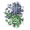
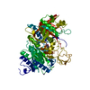
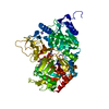
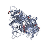
| Title | Crystal structure of beta-ketoacyl synthase from Brucella melitensis in complex with platencin | ||||||
 Components Components | Beta-ketoacyl synthase | ||||||
 Keywords Keywords | Transferase/Antibiotic / SSGCID / beta-ketoacyl synthase / platencin / Structural Genomics / Seattle Structural Genomics Center for Infectious Disease / Transferase-Antibiotic complex | ||||||
| Function / homology |  Function and homology information Function and homology informationbeta-ketoacyl-[acyl-carrier-protein] synthase I / 3-oxoacyl-[acyl-carrier-protein] synthase activity / fatty acid biosynthetic process / cytosol Similarity search - Function | ||||||
| Biological species |  Brucella melitensis biovar Abortus (bacteria) Brucella melitensis biovar Abortus (bacteria) | ||||||
| Method |  X-RAY DIFFRACTION / MOLECULAR REPLACEMENT / Resolution: 1.7 Å X-RAY DIFFRACTION / MOLECULAR REPLACEMENT / Resolution: 1.7 Å | ||||||
 Authors Authors | Seattle Structural Genomics Center for Infectious Disease (SSGCID) | ||||||
 Citation Citation | Journal: Proteins / Year: 2020 Title: Structural characterization of beta-ketoacyl ACP synthase I bound to platencin and fragment screening molecules at two substrate binding sites. Authors: Patterson, E.I. / Nanson, J.D. / Abendroth, J. / Bryan, C. / Sankaran, B. / Myler, P.J. / Forwood, J.K. | ||||||
| History |
|
- Structure visualization
| Structure viewer | Molecule: MolmilJmol/JSmol |
|---|
- Downloads & links
-Download
| PDBx/mmCIF format | 4jv3.cif.gz | 175.2 KB | Display | PDBx/mmCIF format |
|---|---|---|---|---|
| PDB format | pdb4jv3.ent.gz | 135 KB | Display | PDB format |
| PDBx/mmJSON format | 4jv3.json.gz | Tree view | PDBx/mmJSON format | |
| Others |  Other downloads Other downloads |
-Validation report
| Arichive directory | https://data.pdbj.org/pub/pdb/validation_reports/jv/4jv3ftp://data.pdbj.org/pub/pdb/validation_reports/jv/4jv3 | HTTPS FTP |
|---|
-Related structure data
| Related structure data | 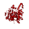 3lrfSC 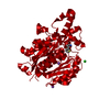 3mqdC 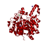 3u0eC 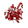 3u0fC S: Starting model for refinement C: citing same article ( |
|---|---|
| Similar structure data | |
| Other databases |


-Links
 PDBj
PDBj

- Assembly
| Deposited unit | 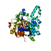
| ||||||||
|---|---|---|---|---|---|---|---|---|---|
| 1 | 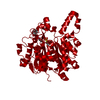
| ||||||||
| Unit cell |
| ||||||||
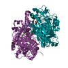
| Details | biological unit is a dimer generated from the monomer in the asymmetric unit by the operation: -X+1, Y, -Z+1 |
-Components
| #1: Protein | Mass: 45723.336 Da / Num. of mol.: 1 Source method: isolated from a genetically manipulated source Source: (gene. exp.) Brucella melitensis biovar Abortus (bacteria)Strain: 2308 / Gene: fabB, BAB1_2173 / Plasmid: AVA0421 / Production host: References: UniProt: Q2YQQ9, beta-ketoacyl-[acyl-carrier-protein] synthase I |
|---|---|
| #2: Chemical | ChemComp-N32 / 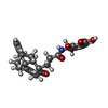  Mass: 425.474 Da / Num. of mol.: 1 / Source method: obtained synthetically / Formula: C24H27NO6 Mass: 425.474 Da / Num. of mol.: 1 / Source method: obtained synthetically / Formula: C24H27NO6 |
| #3: Water | ChemComp-HOH /  Mass: 18.015 Da / Num. of mol.: 433 / Source method: isolated from a natural source / Formula: H2O Mass: 18.015 Da / Num. of mol.: 433 / Source method: isolated from a natural source / Formula: H2O |
-Experimental details
-Experiment
| Experiment | Method: X-RAY DIFFRACTION / Number of used crystals: 1 |
|---|
- Sample preparation
| Crystal | Density Matthews: 2.3 Å3/Da / Density % sol: 46.41 % |
|---|---|
| Crystal grow | Temperature: 290 K / Method: vapor diffusion, sitting drop / pH: 8.5 Details: MD PACT screen, H2: 20% PEG 3350, 100mM BisTrisPropane pH 8.5, 200mM Na-bromide; tray 233841 f11, BrabA.00113.a at 23 mg/ml, 2.5mM platencin, VAPOR DIFFUSION, SITTING DROP, temperature 290K |
-Data collection
| Diffraction | Mean temperature: 100 K | |||||||||||||||||||||||||||||||||||||||||||||||||||||||||||||||||||||||||||||||||||||||||||||||||||||||||||||||||||||||||||||||||||||||||||||||||||
|---|---|---|---|---|---|---|---|---|---|---|---|---|---|---|---|---|---|---|---|---|---|---|---|---|---|---|---|---|---|---|---|---|---|---|---|---|---|---|---|---|---|---|---|---|---|---|---|---|---|---|---|---|---|---|---|---|---|---|---|---|---|---|---|---|---|---|---|---|---|---|---|---|---|---|---|---|---|---|---|---|---|---|---|---|---|---|---|---|---|---|---|---|---|---|---|---|---|---|---|---|---|---|---|---|---|---|---|---|---|---|---|---|---|---|---|---|---|---|---|---|---|---|---|---|---|---|---|---|---|---|---|---|---|---|---|---|---|---|---|---|---|---|---|---|---|---|---|---|
| Diffraction source | Source: ROTATING ANODE / Type: RIGAKU / Wavelength: 1.5418 Å | |||||||||||||||||||||||||||||||||||||||||||||||||||||||||||||||||||||||||||||||||||||||||||||||||||||||||||||||||||||||||||||||||||||||||||||||||||
| Detector | Type: RIGAKU SATURN 944+ / Detector: CCD / Date: Feb 7, 2013 / Details: Rigaku VariMax | |||||||||||||||||||||||||||||||||||||||||||||||||||||||||||||||||||||||||||||||||||||||||||||||||||||||||||||||||||||||||||||||||||||||||||||||||||
| Radiation | Monochromator: RIGAKU VARIMAX / Protocol: SINGLE WAVELENGTH / Monochromatic (M) / Laue (L): M / Scattering type: x-ray | |||||||||||||||||||||||||||||||||||||||||||||||||||||||||||||||||||||||||||||||||||||||||||||||||||||||||||||||||||||||||||||||||||||||||||||||||||
| Radiation wavelength | Wavelength: 1.5418 Å / Relative weight: 1 | |||||||||||||||||||||||||||||||||||||||||||||||||||||||||||||||||||||||||||||||||||||||||||||||||||||||||||||||||||||||||||||||||||||||||||||||||||
| Reflection | Resolution: 1.7→50 Å / Num. all: 45545 / Num. obs: 44869 / % possible obs: 98.5 % / Observed criterion σ(F): 0 / Observed criterion σ(I): -3 / Redundancy: 11.2 % / Biso Wilson estimate: 20.929 Å2 / Rmerge(I) obs: 0.033 / Net I/σ(I): 47.79 | |||||||||||||||||||||||||||||||||||||||||||||||||||||||||||||||||||||||||||||||||||||||||||||||||||||||||||||||||||||||||||||||||||||||||||||||||||
| Reflection shell |
|
- Processing
| Software |
| |||||||||||||||||||||||||||||||||||||||||||||||||||||||||||||||||||||||||||
|---|---|---|---|---|---|---|---|---|---|---|---|---|---|---|---|---|---|---|---|---|---|---|---|---|---|---|---|---|---|---|---|---|---|---|---|---|---|---|---|---|---|---|---|---|---|---|---|---|---|---|---|---|---|---|---|---|---|---|---|---|---|---|---|---|---|---|---|---|---|---|---|---|---|---|---|---|
| Refinement | Method to determine structure: MOLECULAR REPLACEMENT Starting model: pdb entry 3LRF Resolution: 1.7→42.28 Å / Cor.coef. Fo:Fc: 0.969 / Cor.coef. Fo:Fc free: 0.959 / WRfactor Rfree: 0.159 / WRfactor Rwork: 0.1335 / Occupancy max: 1 / Occupancy min: 0.4 / FOM work R set: 0.9088 / SU B: 2.772 / SU ML: 0.048 / SU R Cruickshank DPI: 0.0868 / SU Rfree: 0.0843 / Isotropic thermal model: isotropic, TLS / Cross valid method: THROUGHOUT / σ(F): 0 / ESU R: 0.087 / ESU R Free: 0.084 / Stereochemistry target values: MAXIMUM LIKELIHOOD Details: HYDROGENS HAVE BEEN ADDED IN THE RIDING POSITIONS U VALUES : WITH TLS ADDED
| |||||||||||||||||||||||||||||||||||||||||||||||||||||||||||||||||||||||||||
| Solvent computation | Ion probe radii: 0.8 Å / Shrinkage radii: 0.8 Å / VDW probe radii: 1.2 Å / Solvent model: MASK | |||||||||||||||||||||||||||||||||||||||||||||||||||||||||||||||||||||||||||
| Displacement parameters | Biso max: 49.4 Å2 / Biso mean: 16.929 Å2 / Biso min: 3.56 Å2
| |||||||||||||||||||||||||||||||||||||||||||||||||||||||||||||||||||||||||||
| Refinement step | Cycle: LAST / Resolution: 1.7→42.28 Å
| |||||||||||||||||||||||||||||||||||||||||||||||||||||||||||||||||||||||||||
| Refine LS restraints |
| |||||||||||||||||||||||||||||||||||||||||||||||||||||||||||||||||||||||||||
| LS refinement shell | Resolution: 1.7→1.744 Å / Total num. of bins used: 20
| |||||||||||||||||||||||||||||||||||||||||||||||||||||||||||||||||||||||||||
| Refinement TLS params. | Method: refined / Origin x: 25.63 Å / Origin y: 2.007 Å / Origin z: 17.864 Å
|
