 Movie
Movie Controller

[English] 日本語
 Yorodumi
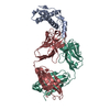
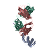
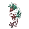
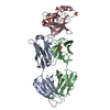
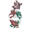
- PDB-6d2p: Crystal structure of IOMA-class CLK31 Fab from an HIV-1 naive don... -
+ Open data

- Basic information
| Entry | Database: PDB / ID: 6d2p | ||||||
|---|---|---|---|---|---|---|---|
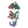
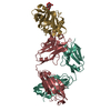
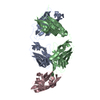
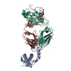
| Title | Crystal structure of IOMA-class CLK31 Fab from an HIV-1 naive donor in complex with a germline-targeting gp120 engineered outer domain eOD-GT8 at 2.6 A | ||||||
 Components Components |
| ||||||
 Keywords Keywords | IMMUNE SYSTEM / HIV-1 gp120 / engineered outer domain (eOD) / germline targeting / CD4 binding site / IOMA-class naive human germline antibody / viral-protein complex / healthy HIV-1 negative donor | ||||||
| Function / homology | Immunoglobulins / Immunoglobulin-like / Sandwich / Mainly Beta Function and homology information Function and homology information | ||||||
| Biological species |  Homo sapiens (human) Homo sapiens (human) | ||||||
| Method |  X-RAY DIFFRACTION / SYNCHROTRON / MOLECULAR REPLACEMENT / Resolution: 2.6 Å X-RAY DIFFRACTION / SYNCHROTRON / MOLECULAR REPLACEMENT / Resolution: 2.6 Å | ||||||
 Authors Authors | Sarkar, A. / Wilson, I.A. | ||||||
 Citation Citation | Journal: Sci Transl Med / Year: 2018 Title: The human naive B cell repertoire contains distinct subclasses for a germline-targeting HIV-1 vaccine immunogen. Authors: Havenar-Daughton, C. / Sarkar, A. / Kulp, D.W. / Toy, L. / Hu, X. / Deresa, I. / Kalyuzhniy, O. / Kaushik, K. / Upadhyay, A.A. / Menis, S. / Landais, E. / Cao, L. / Diedrich, J.K. / Kumar, S. ...Authors: Havenar-Daughton, C. / Sarkar, A. / Kulp, D.W. / Toy, L. / Hu, X. / Deresa, I. / Kalyuzhniy, O. / Kaushik, K. / Upadhyay, A.A. / Menis, S. / Landais, E. / Cao, L. / Diedrich, J.K. / Kumar, S. / Schiffner, T. / Reiss, S.M. / Seumois, G. / Yates, J.R. / Paulson, J.C. / Bosinger, S.E. / Wilson, I.A. / Schief, W.R. / Crotty, S. | ||||||
| History |
|
- Structure visualization
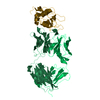
| Structure viewer | Molecule: MolmilJmol/JSmol |
|---|
- Downloads & links
-Download
| PDBx/mmCIF format | 6d2p.cif.gz | 128.7 KB | Display | PDBx/mmCIF format |
|---|---|---|---|---|
| PDB format | pdb6d2p.ent.gz | 97.9 KB | Display | PDB format |
| PDBx/mmJSON format | 6d2p.json.gz | Tree view | PDBx/mmJSON format | |
| Others |  Other downloads Other downloads |
-Validation report
| Arichive directory | https://data.pdbj.org/pub/pdb/validation_reports/d2/6d2pftp://data.pdbj.org/pub/pdb/validation_reports/d2/6d2p | HTTPS FTP |
|---|
-Related structure data
| Related structure data |  5iesS S: Starting model for refinement |
|---|---|
| Similar structure data |


-Links
 PDBj
PDBj

- Assembly
| Deposited unit | 
| ||||||||
|---|---|---|---|---|---|---|---|---|---|
| 1 |
| ||||||||
| Unit cell |
|
-Components
| #1: Protein | Mass: 19889.309 Da / Num. of mol.: 1 Source method: isolated from a genetically manipulated source Source: (gene. exp.) Homo sapiens (human) / Plasmid: pHLsec / Cell line (production host): HEK 293S / Production host: Homo sapiens (human) | ||||
|---|---|---|---|---|---|
| #2: Antibody | Mass: 23697.631 Da / Num. of mol.: 1 Source method: isolated from a genetically manipulated source Source: (gene. exp.) Homo sapiens (human) / Plasmid: pFUSEss-CHIg-hG1 / Cell line (production host): HEK 293 Freestyle / Production host: Homo sapiens (human) | ||||
| #3: Antibody | Mass: 22417.773 Da / Num. of mol.: 1 Source method: isolated from a genetically manipulated source Source: (gene. exp.) Homo sapiens (human) / Plasmid: pFUSEss-CHIg-hG1 / Cell line (production host): HEK 293 Freestyle / Production host: Homo sapiens (human) | ||||
| #4: Chemical | ChemComp-GOL /   Mass: 92.094 Da / Num. of mol.: 5 / Source method: obtained synthetically / Formula: C3H8O3 Mass: 92.094 Da / Num. of mol.: 5 / Source method: obtained synthetically / Formula: C3H8O3#5: Water | ChemComp-HOH / |  Mass: 18.015 Da / Num. of mol.: 113 / Source method: isolated from a natural source / Formula: H2O Mass: 18.015 Da / Num. of mol.: 113 / Source method: isolated from a natural source / Formula: H2OHas protein modification | Y | |
-Experimental details
-Experiment
| Experiment | Method: X-RAY DIFFRACTION / Number of used crystals: 1 |
|---|
- Sample preparation
| Crystal | Density Matthews: 2.7 Å3/Da / Density % sol: 54.48 % |
|---|---|
| Crystal grow | Temperature: 293.15 K / Method: vapor diffusion, sitting drop Details: pH 5.0 of 20% w/v PEG6000, 0.1M citric acid (pH 4.0) |
-Data collection
| Diffraction | Mean temperature: 293 K |
|---|---|
| Diffraction source | Source: SYNCHROTRON / Site: APS  / Beamline: 23-ID-B / Wavelength: 1.0332 Å / Beamline: 23-ID-B / Wavelength: 1.0332 Å |
| Detector | Type: DECTRIS EIGER X 16M / Detector: PIXEL / Date: Oct 28, 2017 |
| Radiation | Protocol: SINGLE WAVELENGTH / Monochromatic (M) / Laue (L): M / Scattering type: x-ray |
| Radiation wavelength | Wavelength: 1.0332 Å / Relative weight: 1 |
| Reflection | Resolution: 2.6→38.26 Å / Num. obs: 21985 / % possible obs: 99.9 % / Redundancy: 10.9 % / CC1/2: 0.92 / Net I/σ(I): 7 |
| Reflection shell | Resolution: 2.6→2.72 Å / Redundancy: 10.8 % / Num. unique obs: 2165 / CC1/2: 0.31 / % possible all: 100 |
- Processing
| Software |
| |||||||||||||||||||||||||||||||||||||||||||||||||||||||||||||||
|---|---|---|---|---|---|---|---|---|---|---|---|---|---|---|---|---|---|---|---|---|---|---|---|---|---|---|---|---|---|---|---|---|---|---|---|---|---|---|---|---|---|---|---|---|---|---|---|---|---|---|---|---|---|---|---|---|---|---|---|---|---|---|---|---|
| Refinement | Method to determine structure: MOLECULAR REPLACEMENT Starting model: 5IES Resolution: 2.6→38.259 Å / SU ML: 0.33 / Cross valid method: FREE R-VALUE / σ(F): 0.31 / Phase error: 26.22 / Stereochemistry target values: ML
| |||||||||||||||||||||||||||||||||||||||||||||||||||||||||||||||
| Solvent computation | Shrinkage radii: 0.9 Å / VDW probe radii: 1.2 Å / Solvent model: FLAT BULK SOLVENT MODEL | |||||||||||||||||||||||||||||||||||||||||||||||||||||||||||||||
| Refinement step | Cycle: LAST / Resolution: 2.6→38.259 Å
| |||||||||||||||||||||||||||||||||||||||||||||||||||||||||||||||
| Refine LS restraints |
| |||||||||||||||||||||||||||||||||||||||||||||||||||||||||||||||
| LS refinement shell |
|
