 Movie
Movie Controller

[English] 日本語
 Yorodumi
- PDB-3rkd: Hepatitis E Virus E2s domain (Genotype I) in complex with a neutr... -
+ Open data

- Basic information
| Entry | Database: PDB / ID: 3rkd | ||||||
|---|---|---|---|---|---|---|---|
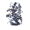
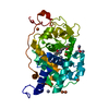
| Title | Hepatitis E Virus E2s domain (Genotype I) in complex with a neutralizing antibody | ||||||
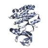
 Components Components |
| ||||||
 Keywords Keywords | VIRAL PROTEIN/IMMUNE SYSTEM / Hepatitis E virus capsid protein / neutralizing antibody / VIRAL PROTEIN-IMMUNE SYSTEM complex | ||||||
| Function / homology |  Function and homology information Function and homology informationT=1 icosahedral viral capsid / host cell endoplasmic reticulum / host cell surface / host cell Golgi apparatus / entry receptor-mediated virion attachment to host cell / symbiont entry into host cell / host cell nucleus / structural molecule activity / RNA binding / extracellular region / identical protein binding Similarity search - Function | ||||||
| Biological species |  Hepatitis E virus Hepatitis E virus | ||||||
| Method |  X-RAY DIFFRACTION / SYNCHROTRON / MOLECULAR REPLACEMENT / Resolution: 1.9 Å X-RAY DIFFRACTION / SYNCHROTRON / MOLECULAR REPLACEMENT / Resolution: 1.9 Å | ||||||
 Authors Authors | Tang, X.H. / Sivaraman, J. | ||||||
 Citation Citation | Journal: Proc.Natl.Acad.Sci.USA / Year: 2011 Title: Structural basis for the neutralization and genotype specificity of hepatitis E virus Authors: Tang, X.H. / Yang, C.Y. / Gu, Y. / Song, C.L. / Zhang, X. / Wang, Y.B. / Zhang, J. / Hew, C.L. / Li, S.W. / Xia, N.S. / Sivaraman, J. | ||||||
| History |
|
- Structure visualization
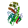
| Structure viewer | Molecule: MolmilJmol/JSmol |
|---|
- Downloads & links
-Download
| PDBx/mmCIF format | 3rkd.cif.gz | 242.4 KB | Display | PDBx/mmCIF format |
|---|---|---|---|---|
| PDB format | pdb3rkd.ent.gz | 192.4 KB | Display | PDB format |
| PDBx/mmJSON format | 3rkd.json.gz | Tree view | PDBx/mmJSON format | |
| Others |  Other downloads Other downloads |
-Validation report
| Arichive directory | https://data.pdbj.org/pub/pdb/validation_reports/rk/3rkdftp://data.pdbj.org/pub/pdb/validation_reports/rk/3rkd | HTTPS FTP |
|---|
-Related structure data
| Related structure data | 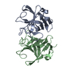 3rkcC  1qblS  3ggqS C: citing same article ( S: Starting model for refinement |
|---|---|
| Similar structure data |


-Links
 PDBj
PDBj


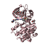
- Assembly
| Deposited unit | 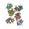
| ||||||||
|---|---|---|---|---|---|---|---|---|---|
| 1 | 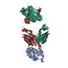
| ||||||||
| 2 | 
| ||||||||
| Unit cell |
|
-Components
| #1: Protein | Mass: 15510.313 Da / Num. of mol.: 2 / Fragment: UNP residues 459-603 / Mutation: Y532H Source method: isolated from a genetically manipulated source Source: (gene. exp.) Hepatitis E virus / Gene: ORF2 / Plasmid: pTO-T7 / Production host:  #2: Antibody | Mass: 23648.018 Da / Num. of mol.: 2 / Source method: isolated from a natural source / Source: (natural) #3: Antibody | Mass: 24476.594 Da / Num. of mol.: 2 / Source method: isolated from a natural source / Source: (natural) #4: Water | ChemComp-HOH / |  Mass: 18.015 Da / Num. of mol.: 761 / Source method: isolated from a natural source / Formula: H2O Mass: 18.015 Da / Num. of mol.: 761 / Source method: isolated from a natural source / Formula: H2OHas protein modification | Y | |
|---|
-Experimental details
-Experiment
| Experiment | Method: X-RAY DIFFRACTION / Number of used crystals: 1 |
|---|
- Sample preparation
| Crystal | Density Matthews: 2.88 Å3/Da / Density % sol: 57.29 % |
|---|---|
| Crystal grow | Temperature: 294 K / Method: vapor diffusion, hanging drop / pH: 7.2 Details: 0.1M HEPEPS, 0.4M KSCN, 0.4M NH4Cl, 18% PEG 3350, 5% w/v n-Dodecyl-beta-D-maltoside, pH 7.2, VAPOR DIFFUSION, HANGING DROP, temperature 294K |
-Data collection
| Diffraction | Mean temperature: 100 K |
|---|---|
| Diffraction source | Source: SYNCHROTRON / Site: NSRRC  / Beamline: BL13B1 / Wavelength: 1 Å / Beamline: BL13B1 / Wavelength: 1 Å |
| Detector | Type: ADSC QUANTUM 315r / Detector: CCD / Date: Nov 17, 2009 |
| Radiation | Monochromator: Si 111 CHANNEL / Protocol: SINGLE WAVELENGTH / Monochromatic (M) / Laue (L): M / Scattering type: x-ray |
| Radiation wavelength | Wavelength: 1 Å / Relative weight: 1 |
| Reflection | Resolution: 1.9→30 Å / Num. obs: 112759 / Redundancy: 6.4 % |
- Processing
| Software |
| ||||||||||||||||||||
|---|---|---|---|---|---|---|---|---|---|---|---|---|---|---|---|---|---|---|---|---|---|
| Refinement | Method to determine structure: MOLECULAR REPLACEMENT Starting model: PDB ENTRY 3GGQ and 1QBL Resolution: 1.9→20 Å / σ(F): 2 / Stereochemistry target values: Engh & Huber
| ||||||||||||||||||||
| Solvent computation | Bsol: 45.6126 Å2 | ||||||||||||||||||||
| Displacement parameters | Biso mean: 26.0299 Å2
| ||||||||||||||||||||
| Refinement step | Cycle: LAST / Resolution: 1.9→20 Å
| ||||||||||||||||||||
| Refine LS restraints |
|
