 Movie
Movie Controller


+ Open data

- Basic information
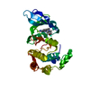
| Entry | Database: PDB / ID: 3rkc | ||||||
|---|---|---|---|---|---|---|---|
| Title | Hepatitis E Virus Capsid Protein E2s Domain (genotype IV) | ||||||
 Components Components | Capsid protein | ||||||
 Keywords Keywords | VIRAL PROTEIN / Viral Capsid Protein | ||||||
| Function / homology | : / Structural protein 2 second domain / : / Structural protein 2 C-terminal domain / viral capsid / identical protein binding / Capsid protein Function and homology information Function and homology information | ||||||
| Biological species |  Hepatitis E virus Hepatitis E virus | ||||||
| Method |  X-RAY DIFFRACTION / MOLECULAR REPLACEMENT / Resolution: 1.79 Å X-RAY DIFFRACTION / MOLECULAR REPLACEMENT / Resolution: 1.79 Å | ||||||
 Authors Authors | Tang, X.H. / Sivaraman, J. | ||||||
 Citation Citation | Journal: Proc.Natl.Acad.Sci.USA / Year: 2011 Title: Structural basis for the neutralization and genotype specificity of hepatitis E virus Authors: Tang, X.H. / Yang, C.Y. / Gu, Y. / Song, C.L. / Zhang, X. / Wang, Y.B. / Zhang, J. / Hew, C.L. / Li, S.W. / Xia, N.S. / Sivaraman, J. | ||||||
| History |
|
- Structure visualization
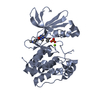
| Structure viewer | Molecule: MolmilJmol/JSmol |
|---|
- Downloads & links
-Download
| PDBx/mmCIF format | 3rkc.cif.gz | 80.8 KB | Display | PDBx/mmCIF format |
|---|---|---|---|---|
| PDB format | pdb3rkc.ent.gz | 59.5 KB | Display | PDB format |
| PDBx/mmJSON format | 3rkc.json.gz | Tree view | PDBx/mmJSON format | |
| Others |  Other downloads Other downloads |
-Validation report
| Arichive directory | https://data.pdbj.org/pub/pdb/validation_reports/rk/3rkcftp://data.pdbj.org/pub/pdb/validation_reports/rk/3rkc | HTTPS FTP |
|---|
-Related structure data
| Related structure data |  3rkdC  3ggqS C: citing same article ( S: Starting model for refinement |
|---|---|
| Similar structure data |


-Links
 PDBj
PDBj- Assembly
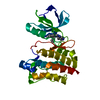
| Deposited unit | 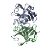
| ||||||||
|---|---|---|---|---|---|---|---|---|---|
| 1 |
| ||||||||
| Unit cell |
|
-Components
| #1: Protein | Mass: 15988.812 Da / Num. of mol.: 2 / Fragment: UNP residues 93-240 Source method: isolated from a genetically manipulated source Source: (gene. exp.) Hepatitis E virus / Plasmid: pTO-T7 / Production host:  #2: Water | ChemComp-HOH / |  Mass: 18.015 Da / Num. of mol.: 567 / Source method: isolated from a natural source / Formula: H2O Mass: 18.015 Da / Num. of mol.: 567 / Source method: isolated from a natural source / Formula: H2O |
|---|
-Experimental details
-Experiment
| Experiment | Method: X-RAY DIFFRACTION / Number of used crystals: 1 |
|---|
- Sample preparation
| Crystal | Density Matthews: 2.21 Å3/Da / Density % sol: 44.29 % |
|---|---|
| Crystal grow | Temperature: 294 K / Method: vapor diffusion, hanging drop / pH: 8 Details: 0.1M Tris, 20% PEG 10000, pH 8.0, VAPOR DIFFUSION, HANGING DROP, temperature 294K |
-Data collection
| Diffraction | Mean temperature: 100 K |
|---|---|
| Diffraction source | Source: ROTATING ANODE / Type: BRUKER AXS MICROSTAR / Wavelength: 1.5418 Å |
| Detector | Type: Bruker Platinum 135 / Detector: CCD / Date: Nov 17, 2009 |
| Radiation | Protocol: SINGLE WAVELENGTH / Monochromatic (M) / Laue (L): M / Scattering type: x-ray |
| Radiation wavelength | Wavelength: 1.5418 Å / Relative weight: 1 |
| Reflection | Resolution: 1.79→50 Å / Num. all: 26223 / Num. obs: 26223 / % possible obs: 100 % / Observed criterion σ(F): 1 / Observed criterion σ(I): 1 / Redundancy: 4.6 % |
- Processing
| Software |
| |||||||||||||||||||||||||
|---|---|---|---|---|---|---|---|---|---|---|---|---|---|---|---|---|---|---|---|---|---|---|---|---|---|---|
| Refinement | Method to determine structure: MOLECULAR REPLACEMENT Starting model: PDB ENTRY 3GGQ Resolution: 1.79→20 Å / σ(F): 2 / Stereochemistry target values: Engh & Huber
| |||||||||||||||||||||||||
| Solvent computation | Bsol: 49.6549 Å2 | |||||||||||||||||||||||||
| Displacement parameters | Biso mean: 13.3327 Å2
| |||||||||||||||||||||||||
| Refinement step | Cycle: LAST / Resolution: 1.79→20 Å
| |||||||||||||||||||||||||
| Refine LS restraints |
|
