 Movie
Movie Controller

[English] 日本語
 Yorodumi
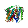
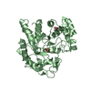
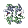
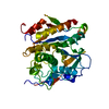
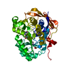
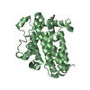
- PDB-6cf3: Ethylene forming enzyme Y306A variant in complex with manganese a... -
+ Open data

- Basic information
| Entry | Database: PDB / ID: 6cf3 | ||||||
|---|---|---|---|---|---|---|---|
| Title | Ethylene forming enzyme Y306A variant in complex with manganese and 2-oxoglutarate | ||||||
 Components Components | 2-oxoglutarate-dependent ethylene/succinate-forming enzyme | ||||||
 Keywords Keywords | OXIDOREDUCTASE / manganese / 2-oxoglutarate / L-arginine / split occupancy / partially occupied / tyrosine to alanine / ethylene glycol / variant induced fold changes | ||||||
| Function / homology |  Function and homology information Function and homology information2-oxoglutarate oxygenase/decarboxylase (ethylene-forming) activity / 2-oxoglutarate dioxygenase (ethene-forming) / 2-oxoglutarate/L-arginine monooxygenase/decarboxylase (succinate-forming) / ethylene biosynthetic process / dioxygenase activity / metal ion binding Similarity search - Function | ||||||
| Biological species |  Pseudomonas savastanoi pv. phaseolicola (bacteria) Pseudomonas savastanoi pv. phaseolicola (bacteria) | ||||||
| Method |  X-RAY DIFFRACTION / SYNCHROTRON / MOLECULAR REPLACEMENT / molecular replacement / Resolution: 1.12 Å X-RAY DIFFRACTION / SYNCHROTRON / MOLECULAR REPLACEMENT / molecular replacement / Resolution: 1.12 Å | ||||||
 Authors Authors | Fellner, M. / Martinez, S. / Hu, J. / Hausinger, R.P. | ||||||
| Funding support |  United States, 1items United States, 1items
| ||||||
 Citation Citation | Journal: Biochemistry / Year: 2024 Title: Structural, Spectroscopic, and Computational Insights from Canavanine-Bound and Two Catalytically Compromised Variants of the Ethylene-Forming Enzyme. Authors: Chatterjee, S. / Fellner, M. / Rankin, J. / Thomas, M.G. / J S Rifayee, S.B. / Christov, C.Z. / Hu, J. / Hausinger, R.P. | ||||||
| History |
|
- Structure visualization
| Structure viewer | Molecule: MolmilJmol/JSmol |
|---|
- Downloads & links
-Download
| PDBx/mmCIF format | 6cf3.cif.gz | 213.4 KB | Display | PDBx/mmCIF format |
|---|---|---|---|---|
| PDB format | pdb6cf3.ent.gz | 171.3 KB | Display | PDB format |
| PDBx/mmJSON format | 6cf3.json.gz | Tree view | PDBx/mmJSON format | |
| Others |  Other downloads Other downloads |
-Validation report
| Arichive directory | https://data.pdbj.org/pub/pdb/validation_reports/cf/6cf3ftp://data.pdbj.org/pub/pdb/validation_reports/cf/6cf3 | HTTPS FTP |
|---|
-Related structure data
| Related structure data |  6cbaC 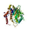 8uc2C  5v2yS S: Starting model for refinement C: citing same article ( |
|---|---|
| Similar structure data |


-Links
 PDBj
PDBj

- Assembly
| Deposited unit | 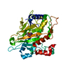
| ||||||||
|---|---|---|---|---|---|---|---|---|---|
| 1 |
| ||||||||
| Unit cell |
|
-Components
| #1: Protein | Mass: 39624.668 Da / Num. of mol.: 1 / Mutation: N-terminal fusion to SH, Y306A Source method: isolated from a genetically manipulated source Source: (gene. exp.) Pseudomonas savastanoi pv. phaseolicola (bacteria)Gene: efe / Plasmid: pET28 / Production host: References: UniProt: P32021, 2-oxoglutarate dioxygenase (ethene-forming), EC: 1.14.11.34 |
|---|---|
| #2: Chemical | ChemComp-MN /   Mass: 54.938 Da / Num. of mol.: 1 / Source method: obtained synthetically / Formula: Mn Mass: 54.938 Da / Num. of mol.: 1 / Source method: obtained synthetically / Formula: Mn |
| #3: Chemical | ChemComp-AKG /   Mass: 146.098 Da / Num. of mol.: 1 / Source method: obtained synthetically / Formula: C5H6O5 Mass: 146.098 Da / Num. of mol.: 1 / Source method: obtained synthetically / Formula: C5H6O5 |
| #4: Chemical | ChemComp-EDO /   Mass: 62.068 Da / Num. of mol.: 1 / Source method: obtained synthetically / Formula: C2H6O2 Mass: 62.068 Da / Num. of mol.: 1 / Source method: obtained synthetically / Formula: C2H6O2 |
| #5: Water | ChemComp-HOH /  Mass: 18.015 Da / Num. of mol.: 351 / Source method: isolated from a natural source / Formula: H2O Mass: 18.015 Da / Num. of mol.: 351 / Source method: isolated from a natural source / Formula: H2O |
-Experimental details
-Experiment
| Experiment | Method: X-RAY DIFFRACTION / Number of used crystals: 1 |
|---|
- Sample preparation
| Crystal | Density Matthews: 2.08 Å3/Da / Density % sol: 40.98 % / Mosaicity: 0.2 ° |
|---|---|
| Crystal grow | Temperature: 277.15 K / Method: vapor diffusion, sitting drop / pH: 6.5 Details: 0.5 ul 64 mg/ml EFE-Y306A (1 mM manganese chloride, 25 mM HEPES pH 8.0, 1 mM TCEP, 3 mM L-Arginine, 3 mM 2-oxoglutarate) was mixed with 0.5 ul reservoir solution. The sitting drop reservoir ...Details: 0.5 ul 64 mg/ml EFE-Y306A (1 mM manganese chloride, 25 mM HEPES pH 8.0, 1 mM TCEP, 3 mM L-Arginine, 3 mM 2-oxoglutarate) was mixed with 0.5 ul reservoir solution. The sitting drop reservoir of 200 ul contained 25% w/v Polyethylene glycol 3,350, 0.1 M Bis-Tris pH 6.5, 0.2 M sodium chloride. The crystal was soaked for five minutes in 25% w/v Ethylene glycol, 75% reservoir solution before freezing it. |
-Data collection
| Diffraction | Mean temperature: 100 K | ||||||||||||||||||||||||
|---|---|---|---|---|---|---|---|---|---|---|---|---|---|---|---|---|---|---|---|---|---|---|---|---|---|
| Diffraction source | Source: SYNCHROTRON / Site: APS / Beamline: 21-ID-D / Wavelength: 0.9762 Å | ||||||||||||||||||||||||
| Detector | Type: DECTRIS EIGER X 9M / Detector: PIXEL / Date: Jun 3, 2017 | ||||||||||||||||||||||||
| Radiation | Protocol: SINGLE WAVELENGTH / Monochromatic (M) / Laue (L): M / Scattering type: x-ray | ||||||||||||||||||||||||
| Radiation wavelength | Wavelength: 0.9762 Å / Relative weight: 1 | ||||||||||||||||||||||||
| Reflection | Resolution: 1.12→44.37 Å / Num. obs: 125336 / % possible obs: 98.2 % / Redundancy: 5.9 % / Biso Wilson estimate: 8.62 Å2 / CC1/2: 0.992 / Rmerge(I) obs: 0.121 / Rpim(I) all: 0.053 / Rrim(I) all: 0.133 / Net I/σ(I): 7.9 | ||||||||||||||||||||||||
| Reflection shell | Diffraction-ID: 1
|
-Phasing
| Phasing | Method: molecular replacement | |||||||||
|---|---|---|---|---|---|---|---|---|---|---|
| Phasing MR |
|
- Processing
| Software |
| |||||||||||||||||||||||||||||||||||||||||||||||||||||||||||||||||||||||||||||||||||||||||||||||||||||||||||||||||||||||||||||||||||||||||||||||||||||||||||||||||||||||||||||||||||||||||||||||||||||||||||||||||||||||||
|---|---|---|---|---|---|---|---|---|---|---|---|---|---|---|---|---|---|---|---|---|---|---|---|---|---|---|---|---|---|---|---|---|---|---|---|---|---|---|---|---|---|---|---|---|---|---|---|---|---|---|---|---|---|---|---|---|---|---|---|---|---|---|---|---|---|---|---|---|---|---|---|---|---|---|---|---|---|---|---|---|---|---|---|---|---|---|---|---|---|---|---|---|---|---|---|---|---|---|---|---|---|---|---|---|---|---|---|---|---|---|---|---|---|---|---|---|---|---|---|---|---|---|---|---|---|---|---|---|---|---|---|---|---|---|---|---|---|---|---|---|---|---|---|---|---|---|---|---|---|---|---|---|---|---|---|---|---|---|---|---|---|---|---|---|---|---|---|---|---|---|---|---|---|---|---|---|---|---|---|---|---|---|---|---|---|---|---|---|---|---|---|---|---|---|---|---|---|---|---|---|---|---|---|---|---|---|---|---|---|---|---|---|---|---|---|---|---|---|
| Refinement | Method to determine structure: MOLECULAR REPLACEMENT Starting model: 5V2Y Resolution: 1.12→44.37 Å / SU ML: 0.1 / Cross valid method: FREE R-VALUE / σ(F): 1.91 / Phase error: 14.97
| |||||||||||||||||||||||||||||||||||||||||||||||||||||||||||||||||||||||||||||||||||||||||||||||||||||||||||||||||||||||||||||||||||||||||||||||||||||||||||||||||||||||||||||||||||||||||||||||||||||||||||||||||||||||||
| Solvent computation | Shrinkage radii: 0.9 Å / VDW probe radii: 1.11 Å | |||||||||||||||||||||||||||||||||||||||||||||||||||||||||||||||||||||||||||||||||||||||||||||||||||||||||||||||||||||||||||||||||||||||||||||||||||||||||||||||||||||||||||||||||||||||||||||||||||||||||||||||||||||||||
| Displacement parameters | Biso max: 49.16 Å2 / Biso mean: 14.2202 Å2 / Biso min: 4.9 Å2 | |||||||||||||||||||||||||||||||||||||||||||||||||||||||||||||||||||||||||||||||||||||||||||||||||||||||||||||||||||||||||||||||||||||||||||||||||||||||||||||||||||||||||||||||||||||||||||||||||||||||||||||||||||||||||
| Refinement step | Cycle: final / Resolution: 1.12→44.37 Å
| |||||||||||||||||||||||||||||||||||||||||||||||||||||||||||||||||||||||||||||||||||||||||||||||||||||||||||||||||||||||||||||||||||||||||||||||||||||||||||||||||||||||||||||||||||||||||||||||||||||||||||||||||||||||||
| LS refinement shell | Refine-ID: X-RAY DIFFRACTION / Rfactor Rfree error: 0 / Total num. of bins used: 30
|
