 Movie
Movie Controller

[English] 日本語
 Yorodumi
- PDB-6bdo: Structure of bacterial type II NADH dehydrogenase from Caldalkali... -
+ Open data

- Basic information
| Entry | Database: PDB / ID: 6bdo | ||||||
|---|---|---|---|---|---|---|---|
| Title | Structure of bacterial type II NADH dehydrogenase from Caldalkalibacillus thermarum complexed with a quinone inhibitor HQNO at 2.8A resolution | ||||||
 Components Components | FAD-dependent pyridine nucleotide-disulfide oxidoreductase | ||||||
 Keywords Keywords | OXIDOREDUCTASE/INHIBITOR / ROSSMANN FOLD / NADH DEHYDROGENASE / OXIDOREDUCTASE / Flavo Protein / MEMBRANE PROTEIN / OXIDOREDUCTASE-INHIBITOR complex | ||||||
| Function / homology |  Function and homology information Function and homology informationaerobic electron transport chain / NAD(P)H dehydrogenase (quinone) activity / nucleotide binding Similarity search - Function | ||||||
| Biological species |   Caldalkalibacillus thermarum TA2.A1 (bacteria) Caldalkalibacillus thermarum TA2.A1 (bacteria) | ||||||
| Method |  X-RAY DIFFRACTION / SYNCHROTRON / MOLECULAR REPLACEMENT / Resolution: 2.8 Å X-RAY DIFFRACTION / SYNCHROTRON / MOLECULAR REPLACEMENT / Resolution: 2.8 Å | ||||||
 Authors Authors | Cook, G.M. / Aragao, D. / Nakatani, Y. | ||||||
| Funding support |  New Zealand, 1items New Zealand, 1items
| ||||||
 Citation Citation | Journal: Biochim. Biophys. Acta / Year: 2018 Title: Structure of the NDH-2 - HQNO inhibited complex provides molecular insight into quinone-binding site inhibitors. Authors: Petri, J. / Shimaki, Y. / Jiao, W. / Bridges, H.R. / Russell, E.R. / Parker, E.J. / Aragao, D. / Cook, G.M. / Nakatani, Y. | ||||||
| History |
|
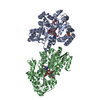
- Structure visualization
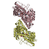
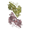
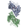
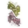
| Structure viewer | Molecule: MolmilJmol/JSmol |
|---|
- Downloads & links
-Download
| PDBx/mmCIF format | 6bdo.cif.gz | 308.1 KB | Display | PDBx/mmCIF format |
|---|---|---|---|---|
| PDB format | pdb6bdo.ent.gz | 246 KB | Display | PDB format |
| PDBx/mmJSON format | 6bdo.json.gz | Tree view | PDBx/mmJSON format | |
| Others |  Other downloads Other downloads |
-Validation report
| Summary document | 6bdo_validation.pdf.gz | 1.8 MB | Display | wwPDB validaton report |
|---|---|---|---|---|
| Full document | 6bdo_full_validation.pdf.gz | 1.9 MB | Display | |
| Data in XML | 6bdo_validation.xml.gz | 57.6 KB | Display | |
| Data in CIF | 6bdo_validation.cif.gz | 75.6 KB | Display | |
| Arichive directory | https://data.pdbj.org/pub/pdb/validation_reports/bd/6bdoftp://data.pdbj.org/pub/pdb/validation_reports/bd/6bdo | HTTPS FTP |
-Related structure data
| Related structure data |  5wedS S: Starting model for refinement |
|---|---|
| Similar structure data |


-Links
 PDBj
PDBj


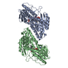
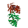
- Assembly
| Deposited unit | 
| ||||||||
|---|---|---|---|---|---|---|---|---|---|
| 1 | 
| ||||||||
| 2 | 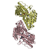
| ||||||||
| Unit cell |
|
-Components
| #1: Protein | Mass: 44571.988 Da / Num. of mol.: 4 Source method: isolated from a genetically manipulated source Source: (gene. exp.) Caldalkalibacillus thermarum TA2.A1 (bacteria)Gene: CathTA2_0279 / Plasmid: PTRC99A / Production host: #2: Chemical | ChemComp-FAD /   Mass: 785.550 Da / Num. of mol.: 4 / Source method: obtained synthetically / Formula: C27H33N9O15P2 / Comment: FAD*YM Mass: 785.550 Da / Num. of mol.: 4 / Source method: obtained synthetically / Formula: C27H33N9O15P2 / Comment: FAD*YM#3: Chemical |   Mass: 259.343 Da / Num. of mol.: 2 / Source method: obtained synthetically / Formula: C16H21NO2 / Feature type: SUBJECT OF INVESTIGATION Mass: 259.343 Da / Num. of mol.: 2 / Source method: obtained synthetically / Formula: C16H21NO2 / Feature type: SUBJECT OF INVESTIGATION#4: Water | ChemComp-HOH / |  Mass: 18.015 Da / Num. of mol.: 43 / Source method: isolated from a natural source / Formula: H2O Mass: 18.015 Da / Num. of mol.: 43 / Source method: isolated from a natural source / Formula: H2O |
|---|
-Experimental details
-Experiment
| Experiment | Method: X-RAY DIFFRACTION / Number of used crystals: 1 |
|---|
- Sample preparation
| Crystal | Density Matthews: 3.04 Å3/Da / Density % sol: 59.51 % |
|---|---|
| Crystal grow | Temperature: 291 K / Method: vapor diffusion, hanging drop / pH: 8.5 Details: 0.1 M BICINE/TRIS BUFFER PH8.5 INCLUDING 10% (V/V) PEG 4000, 25% (V/V) ETHYLENE GLYCOL, 75 MM D,L-LYSINE, 1 MM 2-heptyl-4-hydroxyquinoline-N-oxide, 4% (V/V) dimethyl sulfoxide |
-Data collection
| Diffraction | Mean temperature: 113 K |
|---|---|
| Diffraction source | Source: SYNCHROTRON / Site: Australian Synchrotron  / Beamline: MX2 / Wavelength: 0.9537 Å / Beamline: MX2 / Wavelength: 0.9537 Å |
| Detector | Type: ADSC QUANTUM 315r / Detector: CCD / Date: Nov 12, 2016 |
| Radiation | Protocol: SINGLE WAVELENGTH / Monochromatic (M) / Laue (L): M / Scattering type: x-ray |
| Radiation wavelength | Wavelength: 0.9537 Å / Relative weight: 1 |
| Reflection | Resolution: 2.8→43.345 Å / Num. obs: 52468 / % possible obs: 99.8 % / Redundancy: 3.6 % / CC1/2: 0.998 / Rmerge(I) obs: 0.072 / Rpim(I) all: 0.069 / Net I/σ(I): 14.5 |
| Reflection shell | Resolution: 2.8→2.89 Å / Redundancy: 3.7 % / Rmerge(I) obs: 0.932 / Mean I/σ(I) obs: 1.7 / Num. unique obs: 16726 / CC1/2: 0.446 / Rpim(I) all: 0.883 / Rsym value: 0.932 / % possible all: 100 |
- Processing
| Software |
| ||||||||||||||||||||||||||||||||||||||||||||||||||||||||||||||||||||||||||||||||||||||||||||||||||||||||||||||||||||||||||||||||||||||||||||
|---|---|---|---|---|---|---|---|---|---|---|---|---|---|---|---|---|---|---|---|---|---|---|---|---|---|---|---|---|---|---|---|---|---|---|---|---|---|---|---|---|---|---|---|---|---|---|---|---|---|---|---|---|---|---|---|---|---|---|---|---|---|---|---|---|---|---|---|---|---|---|---|---|---|---|---|---|---|---|---|---|---|---|---|---|---|---|---|---|---|---|---|---|---|---|---|---|---|---|---|---|---|---|---|---|---|---|---|---|---|---|---|---|---|---|---|---|---|---|---|---|---|---|---|---|---|---|---|---|---|---|---|---|---|---|---|---|---|---|---|---|---|
| Refinement | Method to determine structure: MOLECULAR REPLACEMENT Starting model: 5WED Resolution: 2.8→43.345 Å / SU ML: 0.51 / Cross valid method: FREE R-VALUE / σ(F): 1.34 / Phase error: 31.34
| ||||||||||||||||||||||||||||||||||||||||||||||||||||||||||||||||||||||||||||||||||||||||||||||||||||||||||||||||||||||||||||||||||||||||||||
| Solvent computation | Shrinkage radii: 0.9 Å / VDW probe radii: 1.11 Å | ||||||||||||||||||||||||||||||||||||||||||||||||||||||||||||||||||||||||||||||||||||||||||||||||||||||||||||||||||||||||||||||||||||||||||||
| Refinement step | Cycle: LAST / Resolution: 2.8→43.345 Å
| ||||||||||||||||||||||||||||||||||||||||||||||||||||||||||||||||||||||||||||||||||||||||||||||||||||||||||||||||||||||||||||||||||||||||||||
| Refine LS restraints |
| ||||||||||||||||||||||||||||||||||||||||||||||||||||||||||||||||||||||||||||||||||||||||||||||||||||||||||||||||||||||||||||||||||||||||||||
| LS refinement shell |
|
