 Movie
Movie Controller

[English] 日本語
 Yorodumi
- PDB-6b50: Schistosoma mansoni (Blood Fluke) Sulfotransferase/Oxamniquine Co... -
+ Open data

- Basic information
| Entry | Database: PDB / ID: 6b50 | ||||||
|---|---|---|---|---|---|---|---|
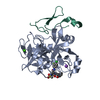
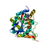
| Title | Schistosoma mansoni (Blood Fluke) Sulfotransferase/Oxamniquine Complex, T157S Mutant | ||||||
 Components Components | Sulfotransferase oxamniquine resistance protein | ||||||
 Keywords Keywords | TRANSFERASE / sulfotransferase / parasite / drug resistance | ||||||
| Function / homology |  Function and homology information Function and homology information | ||||||
| Biological species |  | ||||||
| Method |  X-RAY DIFFRACTION / SYNCHROTRON / MOLECULAR REPLACEMENT / Resolution: 1.87 Å X-RAY DIFFRACTION / SYNCHROTRON / MOLECULAR REPLACEMENT / Resolution: 1.87 Å | ||||||
 Authors Authors | Taylor, A.B. | ||||||
| Funding support |  United States, 1items United States, 1items
| ||||||
 Citation Citation | Journal: Int.J.Parasitol. / Year: 2020 Title: Why does oxamniquine kill Schistosoma mansoni and not S. haematobium and S. japonicum? Authors: Rugel, A.R. / Guzman, M.A. / Taylor, A.B. / Chevalier, F.D. / Tarpley, R.S. / McHardy, S.F. / Cao, X. / Holloway, S.P. / Anderson, T.J.C. / Hart, P.J. / LoVerde, P.T. | ||||||
| History |
|
- Structure visualization
| Structure viewer | Molecule: MolmilJmol/JSmol |
|---|
- Downloads & links
-Download
| PDBx/mmCIF format | 6b50.cif.gz | 72.4 KB | Display | PDBx/mmCIF format |
|---|---|---|---|---|
| PDB format | pdb6b50.ent.gz | 50.4 KB | Display | PDB format |
| PDBx/mmJSON format | 6b50.json.gz | Tree view | PDBx/mmJSON format | |
| Others |  Other downloads Other downloads |
-Validation report
| Summary document | 6b50_validation.pdf.gz | 1000.5 KB | Display | wwPDB validaton report |
|---|---|---|---|---|
| Full document | 6b50_full_validation.pdf.gz | 1001.2 KB | Display | |
| Data in XML | 6b50_validation.xml.gz | 12.8 KB | Display | |
| Data in CIF | 6b50_validation.cif.gz | 17.8 KB | Display | |
| Arichive directory | https://data.pdbj.org/pub/pdb/validation_reports/b5/6b50ftp://data.pdbj.org/pub/pdb/validation_reports/b5/6b50 | HTTPS FTP |
-Related structure data
| Related structure data | 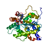 6b4xC  6b4yC  6b4zC  6b51C  6b52C  6b53C  6b54C  5bykS S: Starting model for refinement C: citing same article ( |
|---|---|
| Similar structure data |


-Links
 PDBj
PDBj
- Assembly
| Deposited unit | 
| ||||||||
|---|---|---|---|---|---|---|---|---|---|
| 1 |
| ||||||||
| 2 | 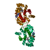
| ||||||||
| Unit cell |
|
-Components
| #1: Protein | Mass: 29962.539 Da / Num. of mol.: 1 / Mutation: T157S Source method: isolated from a genetically manipulated source Source: (gene. exp.)  References: UniProt: G4VLE5, Transferases; Transferring sulfur-containing groups; Sulfotransferases |
|---|---|
| #2: Chemical | ChemComp-A3P /   Type: RNA linking / Mass: 427.201 Da / Num. of mol.: 1 / Source method: obtained synthetically / Formula: C10H15N5O10P2 Type: RNA linking / Mass: 427.201 Da / Num. of mol.: 1 / Source method: obtained synthetically / Formula: C10H15N5O10P2 |
| #3: Chemical | ChemComp-OAQ / {(  Mass: 279.335 Da / Num. of mol.: 1 / Source method: obtained synthetically / Formula: C14H21N3O3 / Feature type: SUBJECT OF INVESTIGATION / Comment: medication*YM Mass: 279.335 Da / Num. of mol.: 1 / Source method: obtained synthetically / Formula: C14H21N3O3 / Feature type: SUBJECT OF INVESTIGATION / Comment: medication*YM |
| #4: Water | ChemComp-HOH /  Mass: 18.015 Da / Num. of mol.: 130 / Source method: isolated from a natural source / Formula: H2O Mass: 18.015 Da / Num. of mol.: 130 / Source method: isolated from a natural source / Formula: H2O |
-Experimental details
-Experiment
| Experiment | Method: X-RAY DIFFRACTION / Number of used crystals: 1 |
|---|
- Sample preparation
| Crystal | Density Matthews: 2.48 Å3/Da / Density % sol: 50.41 % |
|---|---|
| Crystal grow | Temperature: 295 K / Method: vapor diffusion, sitting drop Details: 1.0 M sodium citrate, 0.1 M sodium cacodylate, pH 6.5 |
-Data collection
| Diffraction | Mean temperature: 100 K |
|---|---|
| Diffraction source | Source: SYNCHROTRON / Site: APS / Beamline: 24-ID-E / Wavelength: 0.97918 Å |
| Detector | Type: ADSC QUANTUM 315 / Detector: CCD / Date: Aug 6, 2014 |
| Radiation | Monochromator: single crystal Si(220) side bounce / Protocol: SINGLE WAVELENGTH / Monochromatic (M) / Laue (L): M / Scattering type: x-ray |
| Radiation wavelength | Wavelength: 0.97918 Å / Relative weight: 1 |
| Reflection | Resolution: 1.87→140.19 Å / Num. obs: 25205 / % possible obs: 99 % / Redundancy: 7 % / Biso Wilson estimate: 25.2 Å2 / Rpim(I) all: 0.046 / Rsym value: 0.103 / Net I/σ(I): 15.3 |
| Reflection shell | Resolution: 1.87→1.97 Å / Redundancy: 7.3 % / Mean I/σ(I) obs: 2 / Num. unique obs: 3574 / Rpim(I) all: 0.442 / Rsym value: 1.042 / % possible all: 98 |
- Processing
| Software |
| |||||||||||||||||||||||||||||||||||||||||||||||||||||||||||||||||||||||||||||||||||||||||||||||||||||||||
|---|---|---|---|---|---|---|---|---|---|---|---|---|---|---|---|---|---|---|---|---|---|---|---|---|---|---|---|---|---|---|---|---|---|---|---|---|---|---|---|---|---|---|---|---|---|---|---|---|---|---|---|---|---|---|---|---|---|---|---|---|---|---|---|---|---|---|---|---|---|---|---|---|---|---|---|---|---|---|---|---|---|---|---|---|---|---|---|---|---|---|---|---|---|---|---|---|---|---|---|---|---|---|---|---|---|---|
| Refinement | Method to determine structure: MOLECULAR REPLACEMENT Starting model: PDB entry 5BYK Resolution: 1.87→53.816 Å / SU ML: 0.27 / Cross valid method: THROUGHOUT / σ(F): 1.5 / Phase error: 23.7
| |||||||||||||||||||||||||||||||||||||||||||||||||||||||||||||||||||||||||||||||||||||||||||||||||||||||||
| Solvent computation | Shrinkage radii: 0.9 Å / VDW probe radii: 1.11 Å | |||||||||||||||||||||||||||||||||||||||||||||||||||||||||||||||||||||||||||||||||||||||||||||||||||||||||
| Displacement parameters | Biso mean: 28 Å2 | |||||||||||||||||||||||||||||||||||||||||||||||||||||||||||||||||||||||||||||||||||||||||||||||||||||||||
| Refinement step | Cycle: LAST / Resolution: 1.87→53.816 Å
| |||||||||||||||||||||||||||||||||||||||||||||||||||||||||||||||||||||||||||||||||||||||||||||||||||||||||
| Refine LS restraints |
| |||||||||||||||||||||||||||||||||||||||||||||||||||||||||||||||||||||||||||||||||||||||||||||||||||||||||
| LS refinement shell |
|
