| Entry | Database: PDB / ID: 6ad9
|
|---|
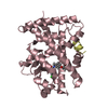
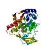
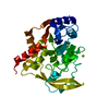
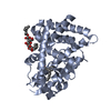
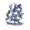
| Title | Crystal Structure of PPARgamma Ligand Binding Domain in complex with dibenzooxepine derivative compound-9 |
|---|
 Components Components | - 12-mer peptide from Peroxisome proliferator-activated receptor gamma coactivator 1-alpha
- Peroxisome proliferator-activated receptor gamma
|
|---|
 Keywords Keywords | NUCLEAR PROTEIN / PPARgamma Ligand Binding Domain Agonist |
|---|
| Function / homology |  Function and homology information Function and homology information
Regulation of MITF-M dependent genes involved in metabolism / positive regulation of fatty acid oxidation / response to muscle activity / Activation of PPARGC1A (PGC-1alpha) by phosphorylation / cellular respiration / lncRNA binding / digestion / temperature homeostasis / response to starvation / intracellular glucose homeostasis ...Regulation of MITF-M dependent genes involved in metabolism / positive regulation of fatty acid oxidation / response to muscle activity / Activation of PPARGC1A (PGC-1alpha) by phosphorylation / cellular respiration / lncRNA binding / digestion / temperature homeostasis / response to starvation / intracellular glucose homeostasis / fatty acid oxidation / response to dietary excess / Transcriptional regulation of brown and beige adipocyte differentiation by EBF2 / Complex I biogenesis / adipose tissue development / Respiratory electron transport / FOXO-mediated transcription of oxidative stress, metabolic and neuronal genes / brown fat cell differentiation / mitochondrial ATP synthesis coupled electron transport / energy homeostasis / transcription coregulator activity / mitochondrial respiratory chain complex I assembly / positive regulation of gluconeogenesis / respiratory electron transport chain / RNA splicing / negative regulation of smooth muscle cell proliferation / SUMOylation of transcription cofactors / nuclear receptor coactivator activity / : / mitochondrion organization / mitochondrial electron transport, NADH to ubiquinone / proton motive force-driven mitochondrial ATP synthesis / Mitochondrial protein degradation / gluconeogenesis / nuclear receptor binding / NADH dehydrogenase (ubiquinone) activity / regulation of circadian rhythm / transcription initiation at RNA polymerase II promoter / PPARA activates gene expression / positive regulation of DNA-binding transcription factor activity / circadian regulation of gene expression / Heme signaling / Transcriptional activation of mitochondrial biogenesis / Transcriptional regulation of white adipocyte differentiation / mitochondrial membrane / aerobic respiration / PML body / chromatin DNA binding / mRNA processing / Regulation of RUNX2 expression and activity / Circadian Clock / positive regulation of cold-induced thermogenesis / cellular response to oxidative stress / DNA-binding transcription factor binding / protein-containing complex assembly / neuron apoptotic process / sequence-specific DNA binding / transcription coactivator activity / negative regulation of neuron apoptotic process / RNA polymerase II-specific DNA-binding transcription factor binding / protein stabilization / mitochondrial inner membrane / ubiquitin protein ligase binding / positive regulation of gene expression / positive regulation of DNA-templated transcription / chromatin / regulation of DNA-templated transcription / positive regulation of transcription by RNA polymerase II / mitochondrion / DNA binding / RNA binding / nucleoplasm / nucleus / cytosolSimilarity search - Function PGC-1alpha, RNA recognition motif / PGC-1 / NADH dehydrogenase 1, beta subcomplex, subunit 6 / NADH:ubiquinone oxidoreductase, NDUFB6/B17 subunit / Retinoid X Receptor / Retinoid X Receptor / RNA recognition motif / RNA recognition motif / Eukaryotic RNA Recognition Motif (RRM) profile. / RNA recognition motif domain ...PGC-1alpha, RNA recognition motif / PGC-1 / NADH dehydrogenase 1, beta subcomplex, subunit 6 / NADH:ubiquinone oxidoreductase, NDUFB6/B17 subunit / Retinoid X Receptor / Retinoid X Receptor / RNA recognition motif / RNA recognition motif / Eukaryotic RNA Recognition Motif (RRM) profile. / RNA recognition motif domain / RNA-binding domain superfamily / Ligand-binding domain of nuclear hormone receptor / Nucleotide-binding alpha-beta plait domain superfamily / Orthogonal Bundle / Mainly AlphaSimilarity search - Domain/homology Chem-KK4 / NADH dehydrogenase [ubiquinone] 1 beta subcomplex subunit 6 / Peroxisome proliferator-activated receptor gamma coactivator 1-alphaSimilarity search - Component |
|---|
| Biological species |  Homo sapiens (human) Homo sapiens (human) |
|---|
| Method |  X-RAY DIFFRACTION / SYNCHROTRON / MOLECULAR REPLACEMENT / molecular replacement / Resolution: 2.2 Å X-RAY DIFFRACTION / SYNCHROTRON / MOLECULAR REPLACEMENT / molecular replacement / Resolution: 2.2 Å |
|---|
 Authors Authors | Takahashi, Y. / Suzuki, M. / Yamamoto, K. / Saito, J. |
|---|
 Citation Citation | Journal: J. Med. Chem. / Year: 2018
Title: Development of Dihydrodibenzooxepine Peroxisome Proliferator-Activated Receptor (PPAR) Gamma Ligands of a Novel Binding Mode as Anticancer Agents: Effective Mimicry of Chiral Structures by Olefinic E/ Z-Isomers.
Authors: Yamamoto, K. / Tamura, T. / Henmi, K. / Kuboyama, T. / Yanagisawa, A. / Matsubara, M. / Takahashi, Y. / Suzuki, M. / Saito, J.I. / Ueno, K. / Shuto, S. |
|---|
| History | | Deposition | Jul 31, 2018 | Deposition site: PDBJ / Processing site: PDBJ |
|---|
| Revision 1.0 | Nov 14, 2018 | Provider: repository / Type: Initial release |
|---|
| Revision 1.1 | Dec 5, 2018 | Group: Data collection / Database references / Category: citation
Item: _citation.journal_volume / _citation.page_first / _citation.page_last |
|---|
| Revision 1.2 | Mar 27, 2024 | Group: Data collection / Database references / Refinement description
Category: chem_comp_atom / chem_comp_bond ...chem_comp_atom / chem_comp_bond / database_2 / refine_hist
Item: _database_2.pdbx_DOI / _database_2.pdbx_database_accession / _refine_hist.d_res_low |
|---|
|
|---|
 Movie
Movie Controller

 Yorodumi
 Open data
 Basic information
 Structure visualization
 Downloads & links
 Other downloads


 PDBj
PDBj


 Assembly
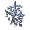


 Mass: 492.568 Da / Num. of mol.: 1 / Source method: obtained synthetically / Formula: C30H28N4O3
 Mass: 18.015 Da / Num. of mol.: 187 / Source method: isolated from a natural source / Formula: H2O
 Sample preparation
 / Beamline: BL-5A / Wavelength: 1 Å
 Processing