[English] 日本語
Yorodumi
- PDB-2baw: Human Nuclear Receptor-Ligand Complex 1 -

+
Open data


ID or keywords:

Loading...

-
Basic information

Entry
Database: PDB / ID: 2baw
TitleHuman Nuclear Receptor-Ligand Complex 1
ComponentsPeroxisome proliferator activated receptor delta
KeywordsTRANSCRIPTION / Nuclear Receptor / Protein-Ligand Complex / PPAR
Function / homology
Function and homology information


fat cell proliferation / positive regulation of fat cell proliferation / keratinocyte migration / linoleic acid binding / positive regulation of skeletal muscle tissue regeneration / axon ensheathment / regulation of skeletal muscle satellite cell proliferation / glucose transmembrane transport / positive regulation of myoblast proliferation / fatty acid catabolic process ...fat cell proliferation / positive regulation of fat cell proliferation / keratinocyte migration / linoleic acid binding / positive regulation of skeletal muscle tissue regeneration / axon ensheathment / regulation of skeletal muscle satellite cell proliferation / glucose transmembrane transport / positive regulation of myoblast proliferation / fatty acid catabolic process / Carnitine metabolism / negative regulation of myoblast differentiation / Regulation of pyruvate dehydrogenase (PDH) complex / Signaling by Retinoic Acid / nuclear steroid receptor activity / positive regulation of fatty acid metabolic process / cell-substrate adhesion / fatty acid beta-oxidation / negative regulation of cholesterol storage / cellular response to nutrient levels / decidualization / keratinocyte proliferation / positive regulation of fat cell differentiation / fatty acid transport / adipose tissue development / energy homeostasis / embryo implantation / hormone-mediated signaling pathway / cholesterol metabolic process / phosphatidylinositol 3-kinase/protein kinase B signal transduction / negative regulation of miRNA transcription / fatty acid metabolic process / generation of precursor metabolites and energy / apoptotic signaling pathway / wound healing / lipid metabolic process / transcription coactivator binding / DNA-binding transcription repressor activity, RNA polymerase II-specific / negative regulation of inflammatory response / Nuclear Receptor transcription pathway / glucose metabolic process / nuclear receptor activity / negative regulation of epithelial cell proliferation / sequence-specific double-stranded DNA binding / cellular response to hypoxia / DNA-binding transcription factor binding / cell population proliferation / positive regulation of phosphatidylinositol 3-kinase/protein kinase B signal transduction / cell differentiation / DNA-binding transcription factor activity, RNA polymerase II-specific / RNA polymerase II cis-regulatory region sequence-specific DNA binding / DNA-binding transcription factor activity / negative regulation of DNA-templated transcription / lipid binding / apoptotic process / positive regulation of gene expression / chromatin / regulation of transcription by RNA polymerase II / positive regulation of DNA-templated transcription / negative regulation of transcription by RNA polymerase II / positive regulation of transcription by RNA polymerase II / DNA binding / zinc ion binding / nucleoplasm / nucleus
Similarity search - Function
Peroxisome proliferator-activated receptor, beta / Peroxisome proliferator-activated receptor / Retinoid X Receptor / Retinoid X Receptor / Nuclear hormone receptor / Nuclear hormones receptors DNA-binding region signature. / Zinc finger, nuclear hormone receptor-type / Zinc finger, C4 type (two domains) / Nuclear hormone receptors DNA-binding domain profile. / c4 zinc finger in nuclear hormone receptors ...Peroxisome proliferator-activated receptor, beta / Peroxisome proliferator-activated receptor / Retinoid X Receptor / Retinoid X Receptor / Nuclear hormone receptor / Nuclear hormones receptors DNA-binding region signature. / Zinc finger, nuclear hormone receptor-type / Zinc finger, C4 type (two domains) / Nuclear hormone receptors DNA-binding domain profile. / c4 zinc finger in nuclear hormone receptors / Nuclear hormone receptor, ligand-binding domain / Nuclear hormone receptor-like domain superfamily / Ligand-binding domain of nuclear hormone receptor / Nuclear receptor (NR) ligand-binding (LBD) domain profile. / Ligand binding domain of hormone receptors / Zinc finger, NHR/GATA-type / Orthogonal Bundle / Mainly Alpha
Similarity search - Domain/homology
heptyl beta-D-glucopyranoside / VACCENIC ACID / Peroxisome proliferator-activated receptor delta
Similarity search - Component
Biological speciesHomo sapiens (human)
MethodX-RAY DIFFRACTION / MOLECULAR REPLACEMENT / Resolution: 2.3 Å
AuthorsFyffe, S.A. / Alphey, M.S. / Buetow, L. / Smith, T.K. / Ferguson, M.A.J. / Sorensen, M.D. / Bjorkling, F. / Hunter, W.N.
CitationJournal: Mol.Cell / Year: 1999
Title: Molecular recognition of fatty acids by peroxisome proliferator-activated receptors.
Authors: Xu, H.E. / Lambert, M.H. / Montana, V.G. / Parks, D.J. / Blanchard, S.G. / Brown, P.J. / Sternbach, D.D. / Lehmann, J.M. / Wisely, G.B. / Willson, T.M. / Kliewer, S.A. / Milburn, M.V.
History
DepositionOct 15, 2005Deposition site: RCSB / Processing site: PDBJ
Revision 1.0Jan 24, 2006Provider: repository / Type: Initial release
Revision 1.1May 1, 2008Group: Version format compliance
Revision 1.2Jul 13, 2011Group: Version format compliance
Revision 1.3Sep 28, 2011Group: Database references
Revision 1.4Jul 29, 2020Group: Data collection / Database references ...Data collection / Database references / Derived calculations / Structure summary
Category: chem_comp / entity ...chem_comp / entity / pdbx_chem_comp_identifier / pdbx_entity_nonpoly / struct_ref_seq_dif / struct_site / struct_site_gen
Item: _chem_comp.mon_nstd_flag / _chem_comp.name ..._chem_comp.mon_nstd_flag / _chem_comp.name / _chem_comp.type / _entity.pdbx_description / _pdbx_entity_nonpoly.name / _struct_ref_seq_dif.details
Description: Carbohydrate remediation / Provider: repository / Type: Remediation
Revision 1.5Mar 13, 2024Group: Data collection / Database references / Structure summary
Category: chem_comp / chem_comp_atom ...chem_comp / chem_comp_atom / chem_comp_bond / database_2
Item: _chem_comp.pdbx_synonyms / _database_2.pdbx_DOI / _database_2.pdbx_database_accession
Remark 0
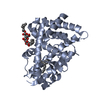
THIS ENTRY 2BAW REFLECTS AN ALTERNATIVE MODELING OF THE STRUCTURAL DATA IN R2GWXSF, ORIGINAL DATA ...THIS ENTRY 2BAW REFLECTS AN ALTERNATIVE MODELING OF THE STRUCTURAL DATA IN R2GWXSF, ORIGINAL DATA DETERMINED BY AUTHOR: H.E.XU,M.H.LAMBERT,V.G.MONTANA, D.J.PARK,S.BLANCHARD,P.BROWN,D.STERNBACH,J.LEHMANN,G.W.BRUCE, T.M.WILLSON,S.A.KLIEWER,M.V.MILBURN

-
Structure visualization

Structure viewerMolecule:
MolmilJmol/JSmol

Downloads & links

-
Assembly

Deposited unit
A: Peroxisome proliferator activated receptor delta
B: Peroxisome proliferator activated receptor delta
hetero molecules


Theoretical massNumber of molelcules
Total (without water)62,2596
Polymers61,1372
Non-polymers1,1224
Water2,900161
1
A: Peroxisome proliferator activated receptor delta
hetero molecules


Theoretical massNumber of molelcules
Total (without water)31,4084
Polymers30,5691
Non-polymers8393
Water181
TypeNameSymmetry operationNumber
identity operation1_555x,y,z1
MethodPISA
2
B: Peroxisome proliferator activated receptor delta
hetero molecules


Theoretical massNumber of molelcules
Total (without water)30,8512
Polymers30,5691
Non-polymers2821
Water181
TypeNameSymmetry operationNumber
identity operation1_555x,y,z1
MethodPISA
Unit cell
Length a, b, c (Å)39.770, 94.220, 96.700
Angle α, β, γ (deg.)90.00, 97.77, 90.00
Int Tables number4
Space group name H-MP1211

-
Components

#1: Protein Peroxisome proliferator activated receptor delta / PPAR-delta / PPAR-beta / Nuclear hormone receptor 1 / NUC1 / NUCI


Mass: 30568.590 Da / Num. of mol.: 2
Source method: isolated from a genetically manipulated source
Source: (gene. exp.) Homo sapiens (human) / Plasmid: PSETA WITH T7 PROMOTER / Species (production host): Escherichia coli / Production host: Escherichia coli BL21(DE3) (bacteria) / Strain (production host): BL21 (DE3) / References: UniProt: Q03181
#2: Sugar ChemComp-B7G / heptyl beta-D-glucopyranoside / HEPTYL-BETA-D-GLUCOPYRANOSIDE / heptyl beta-D-glucoside / heptyl D-glucoside / heptyl glucoside


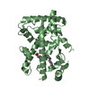
Type: D-saccharide / Mass: 278.342 Da / Num. of mol.: 2 / Source method: obtained synthetically / Formula: C13H26O6
IdentifierTypeProgram
heptyl-b-D-GlucopyranosideIUPAC CARBOHYDRATE SYMBOLPDB-CARE 1.0
#3: Chemical ChemComp-VCA / VACCENIC ACID / (11E)-OCTADEC-11-ENOIC ACID


Mass: 282.461 Da / Num. of mol.: 2 / Source method: obtained synthetically / Formula: C18H34O2
#4: Water ChemComp-HOH / water


Mass: 18.015 Da / Num. of mol.: 161 / Source method: isolated from a natural source / Formula: H2O

-
Experimental details

-
Experiment

ExperimentMethod: X-RAY DIFFRACTION

-
Sample preparation

CrystalDensity Matthews: 2.94 Å3/Da / Density % sol: 58.11 % / Description: AUTHOR USED THE SF DATA FROM ENTRY 2GWX

-
Data collection

RadiationScattering type: x-ray
Radiation wavelengthRelative weight: 1

-
Processing

Software
NameVersionClassificationNB
REFMAC5.2.0005refinement
PDB_EXTRACT1.7data extraction
MOLREPphasing
RefinementMethod to determine structure: MOLECULAR REPLACEMENT / Resolution: 2.3→95.78 Å / Cor.coef. Fo:Fc: 0.887 / Cor.coef. Fo:Fc free: 0.871 / SU B: 7.822 / SU ML: 0.195 / Cross valid method: THROUGHOUT / σ(F): 0 / ESU R: 0.422 / ESU R Free: 0.277 / Stereochemistry target values: MAXIMUM LIKELIHOOD
Details: THIS ENTRY REFLECTS AN ALTERNATIVE MODELING OF X-RAY DATA R2GWXSF. HYDROGENS HAVE BEEN ADDED IN THE RIDING POSITIONS
RfactorNum. reflection% reflectionSelection details
Rfree0.276 1336 5 %RANDOM
Rwork0.245 ---
all0.247 26881 --
obs0.245 25545 85.4 %-
Solvent computationIon probe radii: 0.8 Å / Shrinkage radii: 0.8 Å / VDW probe radii: 1.2 Å / Solvent model: MASK
Displacement parametersBiso mean: 36.706 Å2
Baniso -1Baniso -2Baniso -3
1-0 Å20 Å2-0.03 Å2
2---0.03 Å20 Å2
3---0.03 Å2
Refinement stepCycle: LAST / Resolution: 2.3→95.78 Å
ProteinNucleic acidLigandSolventTotal
Num. atoms3985 0 78 161 4224
Refine LS restraints
Refine-IDTypeDev idealDev ideal targetNumber
X-RAY DIFFRACTIONr_bond_refined_d0.0090.0224139
X-RAY DIFFRACTIONr_angle_refined_deg1.2271.9785570
X-RAY DIFFRACTIONr_dihedral_angle_1_deg5.5255491
X-RAY DIFFRACTIONr_dihedral_angle_2_deg36.33124.033181
X-RAY DIFFRACTIONr_dihedral_angle_3_deg17.39315739
X-RAY DIFFRACTIONr_dihedral_angle_4_deg14.6151522
X-RAY DIFFRACTIONr_chiral_restr0.0860.2647
X-RAY DIFFRACTIONr_gen_planes_refined0.0030.022990
X-RAY DIFFRACTIONr_nbd_refined0.2090.22073
X-RAY DIFFRACTIONr_nbtor_refined0.3020.22857
X-RAY DIFFRACTIONr_xyhbond_nbd_refined0.1560.2192
X-RAY DIFFRACTIONr_symmetry_vdw_refined0.1740.234
X-RAY DIFFRACTIONr_symmetry_hbond_refined0.2160.211
X-RAY DIFFRACTIONr_mcbond_it0.5041.52562
X-RAY DIFFRACTIONr_mcangle_it0.88824011
X-RAY DIFFRACTIONr_scbond_it1.16331743
X-RAY DIFFRACTIONr_scangle_it1.9014.51559
LS refinement shellResolution: 2.3→2.36 Å / Total num. of bins used: 20
RfactorNum. reflection% reflection
Rfree0.287 82 -
Rwork0.222 1503 -
all-1585 -
obs--68.11 %

+
About Yorodumi

-
News

-
Feb 9, 2022. New format data for meta-information of EMDB entries

New format data for meta-information of EMDB entries

  • Version 3 of the EMDB header file is now the official format.
  • The previous official version 1.9 will be removed from the archive.

Related info.:EMDB header

External links:wwPDB to switch to version 3 of the EMDB data model

-
Aug 12, 2020. Covid-19 info

Covid-19 info

URL: https://pdbjlvh1.pdbj.org/emnavi/covid19.php

New page: Covid-19 featured information page in EM Navigator.

Related info.:Covid-19 info / Mar 5, 2020. Novel coronavirus structure data

+
Mar 5, 2020. Novel coronavirus structure data

Novel coronavirus structure data

Related info.:Yorodumi Speices / Aug 12, 2020. Covid-19 info

External links:COVID-19 featured content - PDBj / Molecule of the Month (242):Coronavirus Proteases

+
Jan 31, 2019. EMDB accession codes are about to change! (news from PDBe EMDB page)

EMDB accession codes are about to change! (news from PDBe EMDB page)

  • The allocation of 4 digits for EMDB accession codes will soon come to an end. Whilst these codes will remain in use, new EMDB accession codes will include an additional digit and will expand incrementally as the available range of codes is exhausted. The current 4-digit format prefixed with “EMD-” (i.e. EMD-XXXX) will advance to a 5-digit format (i.e. EMD-XXXXX), and so on. It is currently estimated that the 4-digit codes will be depleted around Spring 2019, at which point the 5-digit format will come into force.
  • The EM Navigator/Yorodumi systems omit the EMD- prefix.

Related info.:Q: What is EMD? / ID/Accession-code notation in Yorodumi/EM Navigator

External links:EMDB Accession Codes are Changing Soon! / Contact to PDBj

+
Jul 12, 2017. Major update of PDB

Major update of PDB

  • wwPDB released updated PDB data conforming to the new PDBx/mmCIF dictionary.
  • This is a major update changing the version number from 4 to 5, and with Remediation, in which all the entries are updated.
  • In this update, many items about electron microscopy experimental information are reorganized (e.g. em_software).
  • Now, EM Navigator and Yorodumi are based on the updated data.

External links:wwPDB Remediation / Enriched Model Files Conforming to OneDep Data Standards Now Available in the PDB FTP Archive

-
Yorodumi

Thousand views of thousand structures

  • Yorodumi is a browser for structure data from EMDB, PDB, SASBDB, etc.
  • This page is also the successor to EM Navigator detail page, and also detail information page/front-end page for Omokage search.
  • The word "yorodu" (or yorozu) is an old Japanese word meaning "ten thousand". "mi" (miru) is to see.

Related info.:EMDB / PDB / SASBDB / Comparison of 3 databanks / Yorodumi Search / Aug 31, 2016. New EM Navigator & Yorodumi / Yorodumi Papers / Jmol/JSmol / Function and homology information / Changes in new EM Navigator and Yorodumi

Read more