 Movie
Movie Controller


+ Open data

- Basic information
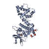
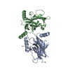
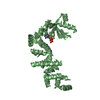
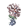
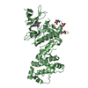
| Entry | Database: PDB / ID: 5xzw | |||||||||||||||
|---|---|---|---|---|---|---|---|---|---|---|---|---|---|---|---|---|
| Title | Crystal structure of Rad53 1-466 | |||||||||||||||
 Components Components | Serine/threonine-protein kinase RAD53 | |||||||||||||||
 Keywords Keywords |  TRANSFERASE / serine/threonine-protein kinase / FHA domain / checkpoint kinase TRANSFERASE / serine/threonine-protein kinase / FHA domain / checkpoint kinase | |||||||||||||||
| Function / homology |  Function and homology information Function and homology informationdeoxyribonucleoside triphosphate biosynthetic process / G2/M DNA damage checkpoint / Chk1/Chk2(Cds1) mediated inactivation of Cyclin B:Cdk1 complex / Ubiquitin Mediated Degradation of Phosphorylated Cdc25A / meiotic recombination checkpoint signaling / Recruitment and ATM-mediated phosphorylation of repair and signaling proteins at DNA double strand breaks / negative regulation of phosphorylation / dual-specificity kinase / DNA replication origin binding / negative regulation of DNA damage checkpoint ...deoxyribonucleoside triphosphate biosynthetic process / G2/M DNA damage checkpoint / Chk1/Chk2(Cds1) mediated inactivation of Cyclin B:Cdk1 complex / Ubiquitin Mediated Degradation of Phosphorylated Cdc25A / meiotic recombination checkpoint signaling / Recruitment and ATM-mediated phosphorylation of repair and signaling proteins at DNA double strand breaks / negative regulation of phosphorylation / dual-specificity kinase / DNA replication origin binding / negative regulation of DNA damage checkpoint / DNA replication initiation / regulation of DNA repair / protein serine/threonine/tyrosine kinase activity / DNA damage checkpoint signaling / protein localization / protein tyrosine kinase activity / protein kinase activity / phosphorylation / DNA repair / protein serine kinase activity / protein serine/threonine kinase activity / ATP binding / nucleus / cytosol / cytoplasmSimilarity search - Function | |||||||||||||||
| Biological species |  Saccharomyces cerevisiae (brewer's yeast) Saccharomyces cerevisiae (brewer's yeast) | |||||||||||||||
| Method | X-RAY DIFFRACTION / SYNCHROTRON / MOLECULAR REPLACEMENT / Resolution: 2.8 Å | |||||||||||||||
 Authors Authors | Weng, J.H. / Tsai, M.D. | |||||||||||||||
| Funding support |  Taiwan, 4items Taiwan, 4items
| |||||||||||||||
 Citation Citation | Journal: Biochemistry / Year: 2017 Title: Phospho-Priming Confers Functionally Relevant Specificities for Rad53 Kinase Autophosphorylation Authors: Chen, E.S. / Weng, J.H. / Chen, Y.H. / Wang, S.C. / Liu, X.X. / Huang, W.C. / Matsui, T. / Kawano, Y. / Liao, J.H. / Lim, L.H. / Bessho, Y. / Huang, K.F. / Wu, W.J. / Tsai, M.D. #1: Journal: To Be PublishedTitle: Phospho-priming Confers Functionally Relevant Specificities for Rad53 Kinase Autophosphorylation Authors: Weng, J.H. / Tsai, M.D. | |||||||||||||||
| History |
|
- Structure visualization
| Structure viewer | Molecule: MolmilJmol/JSmol |
|---|
- Downloads & links
-Download
| PDBx/mmCIF format | 5xzw.cif.gz | 291.9 KB | Display | PDBx/mmCIF format |
|---|---|---|---|---|
| PDB format | pdb5xzw.ent.gz | 247.7 KB | Display | PDB format |
| PDBx/mmJSON format | 5xzw.json.gz | Tree view | PDBx/mmJSON format | |
| Others |  Other downloads Other downloads |
-Validation report
| Arichive directory | https://data.pdbj.org/pub/pdb/validation_reports/xz/5xzwftp://data.pdbj.org/pub/pdb/validation_reports/xz/5xzw | HTTPS FTP |
|---|
-Related structure data


-Links
 PDBj
PDBj


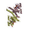
- Assembly
| Deposited unit | 
| ||||||||
|---|---|---|---|---|---|---|---|---|---|
| 1 | 
| ||||||||
| 2 | 
| ||||||||
| 3 |
| ||||||||
| Unit cell |
|
-Components
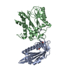
| #1: Protein | Mass: 52506.469 Da / Num. of mol.: 2 / Fragment: UNP RESIDUES 1-466 Source method: isolated from a genetically manipulated source Source: (gene. exp.) Saccharomyces cerevisiae (strain ATCC 204508 / S288c) (yeast)Strain: ATCC 204508 / S288c / Gene: RAD53, MEC2, SAD1, SPK1, YPL153C, P2588 / Production host:  Escherichia coli (E. coli) / References: UniProt: P22216, dual-specificity kinase Escherichia coli (E. coli) / References: UniProt: P22216, dual-specificity kinase#2: Water | ChemComp-HOH / | Water Mass: 18.015 Da / Num. of mol.: 49 / Source method: isolated from a natural source / Formula: H2O Mass: 18.015 Da / Num. of mol.: 49 / Source method: isolated from a natural source / Formula: H2O |
|---|
-Experimental details
-Experiment
| Experiment | Method: X-RAY DIFFRACTION / Number of used crystals: 1 |
|---|
- Sample preparation
| Crystal | Density Matthews: 2.6 Å3/Da / Density % sol: 52.77 % |
|---|---|
| Crystal grow | Temperature: 293 K / Method: vapor diffusion / pH: 6.5 / Details: 100MM CACODYLATE, 0.4M NACL, 1.5M (NH4)2SO4 / PH range: 6.5 |
-Data collection
| Diffraction |
| |||||||||||||||
|---|---|---|---|---|---|---|---|---|---|---|---|---|---|---|---|---|
| Diffraction source |
| |||||||||||||||
| Detector |
| |||||||||||||||
| Radiation |
| |||||||||||||||
| Radiation wavelength |
| |||||||||||||||
| Reflection | Resolution: 2.8→30 Å / Num. obs: 27160 / % possible obs: 98.6 % / Redundancy: 9 % / Biso Wilson estimate: 48.14 Å2 / Rmerge(I) obs: 0.057 / Net I/σ(I): 19.8 | |||||||||||||||
| Reflection shell | Resolution: 2.8→2.9 Å / Redundancy: 9.3 % / Rmerge(I) obs: 0.699 / % possible all: 100 |

- Processing
| Software |
| |||||||||||||||||||||||||||||||||||||||||||||||||||||||||||||||||||||||||||||||||||||||||||||||||||||||||||||||||||||||||||||
|---|---|---|---|---|---|---|---|---|---|---|---|---|---|---|---|---|---|---|---|---|---|---|---|---|---|---|---|---|---|---|---|---|---|---|---|---|---|---|---|---|---|---|---|---|---|---|---|---|---|---|---|---|---|---|---|---|---|---|---|---|---|---|---|---|---|---|---|---|---|---|---|---|---|---|---|---|---|---|---|---|---|---|---|---|---|---|---|---|---|---|---|---|---|---|---|---|---|---|---|---|---|---|---|---|---|---|---|---|---|---|---|---|---|---|---|---|---|---|---|---|---|---|---|---|---|---|
| Refinement | Method to determine structure: MOLECULAR REPLACEMENT / Resolution: 2.8→29.528 Å / SU ML: 0.14 / Cross valid method: FREE R-VALUE / σ(F): 1.34 / Phase error: 28.06 / Stereochemistry target values: ML
| |||||||||||||||||||||||||||||||||||||||||||||||||||||||||||||||||||||||||||||||||||||||||||||||||||||||||||||||||||||||||||||
| Solvent computation | Shrinkage radii: 0.9 Å / VDW probe radii: 1.11 Å / Solvent model: FLAT BULK SOLVENT MODEL | |||||||||||||||||||||||||||||||||||||||||||||||||||||||||||||||||||||||||||||||||||||||||||||||||||||||||||||||||||||||||||||
| Refinement step | Cycle: LAST / Resolution: 2.8→29.528 Å
| |||||||||||||||||||||||||||||||||||||||||||||||||||||||||||||||||||||||||||||||||||||||||||||||||||||||||||||||||||||||||||||
| Refine LS restraints |
| |||||||||||||||||||||||||||||||||||||||||||||||||||||||||||||||||||||||||||||||||||||||||||||||||||||||||||||||||||||||||||||
| LS refinement shell |
| |||||||||||||||||||||||||||||||||||||||||||||||||||||||||||||||||||||||||||||||||||||||||||||||||||||||||||||||||||||||||||||
| Refinement TLS params. | Method: refined / Refine-ID: X-RAY DIFFRACTION
| |||||||||||||||||||||||||||||||||||||||||||||||||||||||||||||||||||||||||||||||||||||||||||||||||||||||||||||||||||||||||||||
| Refinement TLS group |
|
