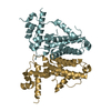
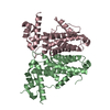
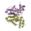
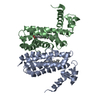
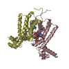
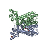
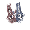
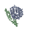
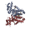
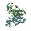
A: Gamma-butyrolactone receptor protein B: Gamma-butyrolactone receptor protein C: Gamma-butyrolactone receptor protein D: Gamma-butyrolactone receptor protein E: Gamma-butyrolactone receptor protein F: Gamma-butyrolactone receptor protein G: Gamma-butyrolactone receptor protein H: Gamma-butyrolactone receptor protein
Protocol: SINGLE WAVELENGTH / Monochromatic (M) / Laue (L): M / Scattering type: x-ray
Radiation wavelength
Wavelength: 0.9686 Å / Relative weight: 1
Reflection
Resolution: 2.58→19.92 Å / Num. obs: 69940 / % possible obs: 99.5 % / Redundancy: 4.1 % / Net I/σ(I): 2.9
Reflection shell
Resolution: 2.58→2.62 Å / % possible obs: 98.4 % / Redundancy: 4.2 % / Rmerge(I) obs: 0.542 / Mean I/σ(I) obs: 2.9 / Rsym value: 0.621
-
Processing
Software
Name
Version
Classification
REFMAC
5.6.0117
refinement
XDS
dataprocessing
Aimless
datascaling
Auto-Rickshaw
phasing
Refinement
Method to determine structure: SAD / Resolution: 2.58→19.92 Å / Cor.coef. Fo:Fc: 0.95 / Cor.coef. Fo:Fc free: 0.916 / SU B: 11.617 / SU ML: 0.241 / Cross valid method: THROUGHOUT / ESU R: 0.488 / ESU R Free: 0.315 / Stereochemistry target values: MAXIMUM LIKELIHOOD / Details: HYDROGENS HAVE BEEN USED IF PRESENT IN THE INPUT
Rfactor
Num. reflection
% reflection
Selection details
Rfree
0.28386
994
1.4 %
RANDOM
Rwork
0.22734
-
-
-
obs
0.22813
69922
99.57 %
-
Solvent computation
Ion probe radii: 0.8 Å / Shrinkage radii: 0.8 Å / VDW probe radii: 1.2 Å / Solvent model: MASK
 Movie
Movie Controller

 Yorodumi
 Open data
 Basic information
 Components
 Keywords
 DNA BINDING PROTEIN /
  Function and homology information
 Authors
 Australia,
  India, 3items
  Citation
 Structure visualization
 Downloads & links
 Other downloads
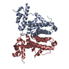


 PDBj
PDBj Assembly
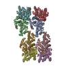

 Mass: 18.015 Da / Num. of mol.: 114 / Source method: isolated from a natural source / Formula: H2O
 Sample preparation
 Processing