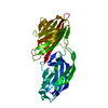
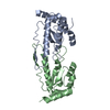
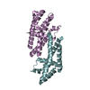
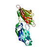
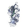
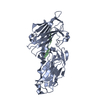
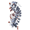
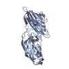
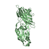
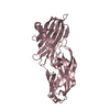
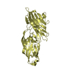
[English] 日本語
Yorodumi
- PDB-5wta: Crystal Structure of Staphylococcus aureus SdrE apo form -

+
Open data


ID or keywords:

Loading...

-
Basic information

Entry
Database: PDB / ID: 5wta
TitleCrystal Structure of Staphylococcus aureus SdrE apo form
ComponentsSerine-aspartate repeat-containing protein E
KeywordsCELL ADHESION / Surface protein / Staphylococcus aureus / Sdr family / complement evasion
Function / homology
Function and homology information


cell adhesion / extracellular region
Similarity search - Function
SD-repeat containing protein, B domain / SdrD B-like domain / Fibrinogen-binding domain 2 / C-terminus of bacterial fibrinogen-binding adhesin / Immunoglobulin-like - #1290 / Immunoglobulin-like - #1280 / SDR-like Ig domain / Bacterial Ig domain / Fibrogen-binding domain 1 / YSIRK type signal peptide ...SD-repeat containing protein, B domain / SdrD B-like domain / Fibrinogen-binding domain 2 / C-terminus of bacterial fibrinogen-binding adhesin / Immunoglobulin-like - #1290 / Immunoglobulin-like - #1280 / SDR-like Ig domain / Bacterial Ig domain / Fibrogen-binding domain 1 / YSIRK type signal peptide / Adhesion domain superfamily / YSIRK Gram-positive signal peptide / LPXTG cell wall anchor motif / Gram-positive cocci surface proteins LPxTG motif profile. / LPXTG cell wall anchor domain / Immunoglobulin-like fold / Immunoglobulin-like / Sandwich / Mainly Beta
Similarity search - Domain/homology
Serine-aspartate repeat-containing protein E
Similarity search - Component
Biological speciesStaphylococcus aureus (bacteria)
MethodX-RAY DIFFRACTION / SYNCHROTRON / MOLECULAR REPLACEMENT / Resolution: 2.3 Å
AuthorsWu, M. / Zhang, Y. / Hang, T. / Wang, C. / Yang, Y. / Zang, J. / Zhang, M. / Zhang, X.
Funding support China, 1items
OrganizationGrant numberCountry
China
CitationJournal: Biochem. J. / Year: 2017
Title: Staphylococcus aureus SdrE captures complement factor H's C-terminus via a novel 'close, dock, lock and latch' mechanism for complement evasion
Authors: Zhang, Y. / Wu, M. / Hang, T. / Wang, C. / Yang, Y. / Pan, W. / Zang, J. / Zhang, M. / Zhang, X.
History
DepositionDec 10, 2016Deposition site: PDBJ / Processing site: PDBJ
Revision 1.0Jul 19, 2017Provider: repository / Type: Initial release
Revision 1.1Nov 8, 2023Group: Data collection / Database references / Refinement description
Category: chem_comp_atom / chem_comp_bond ...chem_comp_atom / chem_comp_bond / database_2 / pdbx_initial_refinement_model
Item: _database_2.pdbx_DOI / _database_2.pdbx_database_accession

-
Structure visualization

Structure viewerMolecule:
MolmilJmol/JSmol

Downloads & links

-
Assembly

Deposited unit
A: Serine-aspartate repeat-containing protein E
B: Serine-aspartate repeat-containing protein E
C: Serine-aspartate repeat-containing protein E
D: Serine-aspartate repeat-containing protein E


Theoretical massNumber of molelcules
Total (without water)150,7774
Polymers150,7774
Non-polymers00
Water11,656647
1
A: Serine-aspartate repeat-containing protein E


Theoretical massNumber of molelcules
Total (without water)37,6941
Polymers37,6941
Non-polymers00
Water181
TypeNameSymmetry operationNumber
identity operation1_555x,y,z1
MethodPISA
2
B: Serine-aspartate repeat-containing protein E


Theoretical massNumber of molelcules
Total (without water)37,6941
Polymers37,6941
Non-polymers00
Water181
TypeNameSymmetry operationNumber
identity operation1_555x,y,z1
MethodPISA
3
C: Serine-aspartate repeat-containing protein E


Theoretical massNumber of molelcules
Total (without water)37,6941
Polymers37,6941
Non-polymers00
Water181
TypeNameSymmetry operationNumber
identity operation1_555x,y,z1
MethodPISA
4
D: Serine-aspartate repeat-containing protein E


Theoretical massNumber of molelcules
Total (without water)37,6941
Polymers37,6941
Non-polymers00
Water181
TypeNameSymmetry operationNumber
identity operation1_555x,y,z1
MethodPISA
Unit cell
Length a, b, c (Å)41.810, 61.730, 139.610
Angle α, β, γ (deg.)80.89, 89.83, 73.34
Int Tables number1
Space group name H-MP1

-
Components

#1: Protein
Serine-aspartate repeat-containing protein E


Mass: 37694.152 Da / Num. of mol.: 4 / Fragment: Ligand binding A domain, UNP residues 270-599 / Mutation: I489M
Source method: isolated from a genetically manipulated source
Source: (gene. exp.) Staphylococcus aureus (strain Mu50 / ATCC 700699) (bacteria)
Strain: Mu50 / ATCC 700699 / Gene: sdrE, SAV0563 / Production host: Escherichia coli BL21(DE3) (bacteria) / Strain (production host): BL21(DE3) / References: UniProt: Q932F7
#2: Water ChemComp-HOH / water / Water


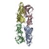
Mass: 18.015 Da / Num. of mol.: 647 / Source method: isolated from a natural source / Formula: H2O

-
Experimental details

-
Experiment

ExperimentMethod: X-RAY DIFFRACTION / Number of used crystals: 1

-
Sample preparation

CrystalDensity Matthews: 2.38 Å3/Da / Density % sol: 48.28 %
Crystal growTemperature: 277 K / Method: vapor diffusion, sitting drop
Details: 0.1M Citric acid, pH 3.5, 16% PEG 3350 (w/v), 2% MPD (v/v)

-
Data collection

DiffractionMean temperature: 100 K
Diffraction sourceSource: SYNCHROTRON / Site: SSRF / Beamline: BL17U / Wavelength: 0.9791 Å
DetectorType: ADSC QUANTUM 315r / Detector: CCD / Date: Jun 12, 2015
RadiationProtocol: SINGLE WAVELENGTH / Monochromatic (M) / Laue (L): M / Scattering type: x-ray
Radiation wavelengthWavelength: 0.9791 Å / Relative weight: 1
ReflectionResolution: 2.3→40.01 Å / Num. obs: 55992 / % possible obs: 95.5 % / Redundancy: 2.2 % / Rsym value: 0.08 / Net I/σ(I): 7.8
Reflection shellResolution: 2.3→2.42 Å / Redundancy: 2.3 % / Mean I/σ(I) obs: 3.4 / Num. unique obs: 8137 / Rsym value: 0.26 / % possible all: 95.2

-
Processing

Software
NameVersionClassification
REFMAC5.7.0032refinement
iMOSFLMdata reduction
SCALAdata scaling
PHASERphasing
RefinementMethod to determine structure: MOLECULAR REPLACEMENT
Starting model: 1R17
Resolution: 2.3→38.93 Å / Cor.coef. Fo:Fc: 0.93 / Cor.coef. Fo:Fc free: 0.882 / Cross valid method: THROUGHOUT / ESU R: 0.454 / ESU R Free: 0.267 / Stereochemistry target values: MAXIMUM LIKELIHOOD / Details: HYDROGENS HAVE BEEN ADDED IN THE RIDING POSITIONS
RfactorNum. reflection% reflectionSelection details
Rfree0.25171 2821 5 %RANDOM
Rwork0.19409 ---
obs0.19693 53169 95.5 %-
Solvent computationIon probe radii: 0.8 Å / Shrinkage radii: 0.8 Å / VDW probe radii: 1.2 Å / Solvent model: MASK
Displacement parametersBiso mean: 22.278 Å2
Baniso -1Baniso -2Baniso -3
1-0.75 Å20.05 Å20.4 Å2
2---0.08 Å2-0.88 Å2
3----0.78 Å2
Refinement stepCycle: 1 / Resolution: 2.3→38.93 Å
ProteinNucleic acidLigandSolventTotal
Num. atoms9900 0 0 647 10547
Refine LS restraints
Refine-IDTypeDev idealDev ideal targetNumber
X-RAY DIFFRACTIONr_bond_refined_d0.0140.0210082
X-RAY DIFFRACTIONr_bond_other_d00.029292
X-RAY DIFFRACTIONr_angle_refined_deg1.4091.94713693
X-RAY DIFFRACTIONr_angle_other_deg3.703321506
X-RAY DIFFRACTIONr_dihedral_angle_1_deg6.27551262
X-RAY DIFFRACTIONr_dihedral_angle_2_deg39.97626.978503
X-RAY DIFFRACTIONr_dihedral_angle_3_deg16.292151766
X-RAY DIFFRACTIONr_dihedral_angle_4_deg13.9751512
X-RAY DIFFRACTIONr_chiral_restr0.0840.21570
X-RAY DIFFRACTIONr_gen_planes_refined0.0050.0211678
X-RAY DIFFRACTIONr_gen_planes_other0.0090.022178
X-RAY DIFFRACTIONr_nbd_refined
X-RAY DIFFRACTIONr_nbd_other
X-RAY DIFFRACTIONr_nbtor_refined
X-RAY DIFFRACTIONr_nbtor_other
X-RAY DIFFRACTIONr_xyhbond_nbd_refined
X-RAY DIFFRACTIONr_xyhbond_nbd_other
X-RAY DIFFRACTIONr_metal_ion_refined
X-RAY DIFFRACTIONr_metal_ion_other
X-RAY DIFFRACTIONr_symmetry_vdw_refined
X-RAY DIFFRACTIONr_symmetry_vdw_other
X-RAY DIFFRACTIONr_symmetry_hbond_refined
X-RAY DIFFRACTIONr_symmetry_hbond_other
X-RAY DIFFRACTIONr_symmetry_metal_ion_refined
X-RAY DIFFRACTIONr_symmetry_metal_ion_other
X-RAY DIFFRACTIONr_mcbond_it1.7132.1375036
X-RAY DIFFRACTIONr_mcbond_other1.7122.1375035
X-RAY DIFFRACTIONr_mcangle_it2.6783.2016290
X-RAY DIFFRACTIONr_mcangle_other2.6783.2016291
X-RAY DIFFRACTIONr_scbond_it2.3672.3155046
X-RAY DIFFRACTIONr_scbond_other2.3672.3155046
X-RAY DIFFRACTIONr_scangle_it
X-RAY DIFFRACTIONr_scangle_other3.6323.3957400
X-RAY DIFFRACTIONr_long_range_B_refined5.20917.29711344
X-RAY DIFFRACTIONr_long_range_B_other5.13317.22711224
X-RAY DIFFRACTIONr_rigid_bond_restr
X-RAY DIFFRACTIONr_sphericity_free
X-RAY DIFFRACTIONr_sphericity_bonded
LS refinement shellResolution: 2.3→2.36 Å / Total num. of bins used: 20
RfactorNum. reflection% reflection
Rfree0.29 219 -
Rwork0.23 3928 -
obs--95.09 %

+
About Yorodumi

-
News

-
Feb 9, 2022. New format data for meta-information of EMDB entries

New format data for meta-information of EMDB entries

  • Version 3 of the EMDB header file is now the official format.
  • The previous official version 1.9 will be removed from the archive.

Related info.:EMDB header

External links:wwPDB to switch to version 3 of the EMDB data model

-
Aug 12, 2020. Covid-19 info

Covid-19 info

URL: https://pdbj.org/emnavi/covid19.php

New page: Covid-19 featured information page in EM Navigator.

Related info.:Covid-19 info / Mar 5, 2020. Novel coronavirus structure data

+
Mar 5, 2020. Novel coronavirus structure data

Novel coronavirus structure data

Related info.:Yorodumi Speices / Aug 12, 2020. Covid-19 info

External links:COVID-19 featured content - PDBj / Molecule of the Month (242):Coronavirus Proteases

+
Jan 31, 2019. EMDB accession codes are about to change! (news from PDBe EMDB page)

EMDB accession codes are about to change! (news from PDBe EMDB page)

  • The allocation of 4 digits for EMDB accession codes will soon come to an end. Whilst these codes will remain in use, new EMDB accession codes will include an additional digit and will expand incrementally as the available range of codes is exhausted. The current 4-digit format prefixed with “EMD-” (i.e. EMD-XXXX) will advance to a 5-digit format (i.e. EMD-XXXXX), and so on. It is currently estimated that the 4-digit codes will be depleted around Spring 2019, at which point the 5-digit format will come into force.
  • The EM Navigator/Yorodumi systems omit the EMD- prefix.

Related info.:Q: What is EMD? / ID/Accession-code notation in Yorodumi/EM Navigator

External links:EMDB Accession Codes are Changing Soon! / Contact to PDBj

+
Jul 12, 2017. Major update of PDB

Major update of PDB

  • wwPDB released updated PDB data conforming to the new PDBx/mmCIF dictionary.
  • This is a major update changing the version number from 4 to 5, and with Remediation, in which all the entries are updated.
  • In this update, many items about electron microscopy experimental information are reorganized (e.g. em_software).
  • Now, EM Navigator and Yorodumi are based on the updated data.

External links:wwPDB Remediation / Enriched Model Files Conforming to OneDep Data Standards Now Available in the PDB FTP Archive

-
Yorodumi

Thousand views of thousand structures

  • Yorodumi is a browser for structure data from EMDB, PDB, SASBDB, etc.
  • This page is also the successor to EM Navigator detail page, and also detail information page/front-end page for Omokage search.
  • The word "yorodu" (or yorozu) is an old Japanese word meaning "ten thousand". "mi" (miru) is to see.

Related info.:EMDB / PDB / SASBDB / Comparison of 3 databanks / Yorodumi Search / Aug 31, 2016. New EM Navigator & Yorodumi / Yorodumi Papers / Jmol/JSmol / Function and homology information / Changes in new EM Navigator and Yorodumi

Read more