 Movie
Movie Controller


+ Open data

- Basic information
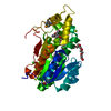
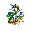
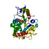
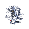
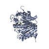
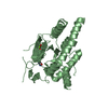
| Entry | Database: PDB / ID: 5wdh | |||||||||
|---|---|---|---|---|---|---|---|---|---|---|
| Title | Motor domain of human kinesin family member C1 | |||||||||
 Components Components | Kinesin-like protein KIFC1 | |||||||||
 Keywords Keywords | MOTOR PROTEIN / kinesin / structural genomics consortium / motor domain / adp / SGC | |||||||||
| Function / homology |  Function and homology information Function and homology informationKinesins / kinesin complex / microtubule motor activity / COPI-dependent Golgi-to-ER retrograde traffic / microtubule-based movement / mitotic metaphase chromosome alignment / microtubule organizing center / mitotic sister chromatid segregation / mitotic spindle assembly / mitotic spindle ...Kinesins / kinesin complex / microtubule motor activity / COPI-dependent Golgi-to-ER retrograde traffic / microtubule-based movement / mitotic metaphase chromosome alignment / microtubule organizing center / mitotic sister chromatid segregation / mitotic spindle assembly / mitotic spindle / microtubule binding / microtubule / early endosome / cell division / centrosome / ATP hydrolysis activity / ATP binding / membrane / nucleus Similarity search - Function | |||||||||
| Biological species |  Homo sapiens (human) Homo sapiens (human) | |||||||||
| Method |  X-RAY DIFFRACTION / SYNCHROTRON / MOLECULAR REPLACEMENT / Resolution: 2.248 Å X-RAY DIFFRACTION / SYNCHROTRON / MOLECULAR REPLACEMENT / Resolution: 2.248 Å | |||||||||
 Authors Authors | Zhu, H. / Tempel, W. / He, H. / Shen, Y. / Wang, J. / Brothers, G. / Landry, R. / Arrowsmith, C.H. / Edwards, A.M. / Park, H. / Structural Genomics Consortium (SGC) | |||||||||
 Citation Citation | Journal: Sci Rep / Year: 2017 Title: Structural basis of small molecule ATPase inhibition of a human mitotic kinesin motor protein. Authors: Park, H.W. / Ma, Z. / Zhu, H. / Jiang, S. / Robinson, R.C. / Endow, S.A. | |||||||||
| History |
|
- Structure visualization
| Structure viewer | Molecule: MolmilJmol/JSmol |
|---|
- Downloads & links
-Download
| PDBx/mmCIF format | 5wdh.cif.gz | 131.4 KB | Display | PDBx/mmCIF format |
|---|---|---|---|---|
| PDB format | pdb5wdh.ent.gz | 98.5 KB | Display | PDB format |
| PDBx/mmJSON format | 5wdh.json.gz | Tree view | PDBx/mmJSON format | |
| Others |  Other downloads Other downloads |
-Validation report
| Arichive directory | https://data.pdbj.org/pub/pdb/validation_reports/wd/5wdhftp://data.pdbj.org/pub/pdb/validation_reports/wd/5wdh | HTTPS FTP |
|---|
-Related structure data
| Related structure data | 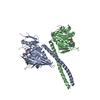 5w3dC  5wdeC 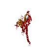 2ncdS S: Starting model for refinement C: citing same article ( |
|---|---|
| Similar structure data |


-Links
 PDBj
PDBj


- Assembly
| Deposited unit | 
| ||||||||
|---|---|---|---|---|---|---|---|---|---|
| 1 |
| ||||||||
| Unit cell |
|
-Components
| #1: Protein | Mass: 40578.613 Da / Num. of mol.: 1 / Fragment: motor domain (UNP residues 307-663) Source method: isolated from a genetically manipulated source Source: (gene. exp.) Homo sapiens (human) / Gene: KIFC1, HSET, KNSL2 / Plasmid: pFBOH-LIC / Cell line (production host): HighFive / Production host:  Trichoplusia ni (cabbage looper) / References: UniProt: Q9BW19 Trichoplusia ni (cabbage looper) / References: UniProt: Q9BW19 | ||
|---|---|---|---|
| #2: Chemical | ChemComp-ADP /   Mass: 427.201 Da / Num. of mol.: 1 / Source method: obtained synthetically / Formula: C10H15N5O10P2 / Comment: ADP, energy-carrying molecule*YM Mass: 427.201 Da / Num. of mol.: 1 / Source method: obtained synthetically / Formula: C10H15N5O10P2 / Comment: ADP, energy-carrying molecule*YM | ||
| #3: Chemical | ChemComp-MG /   Mass: 24.305 Da / Num. of mol.: 1 / Source method: obtained synthetically / Formula: Mg Mass: 24.305 Da / Num. of mol.: 1 / Source method: obtained synthetically / Formula: Mg | ||
| #4: Chemical | ChemComp-UNX /   Num. of mol.: 6 / Source method: obtained synthetically Num. of mol.: 6 / Source method: obtained synthetically#5: Water | ChemComp-HOH / |  Mass: 18.015 Da / Num. of mol.: 4 / Source method: isolated from a natural source / Formula: H2O Mass: 18.015 Da / Num. of mol.: 4 / Source method: isolated from a natural source / Formula: H2O |
-Experimental details
-Experiment
| Experiment | Method: X-RAY DIFFRACTION / Number of used crystals: 1 |
|---|
- Sample preparation
| Crystal | Density Matthews: 3.03 Å3/Da / Density % sol: 59.43 % |
|---|---|
| Crystal grow | Temperature: 291 K / Method: vapor diffusion / pH: 7.5 Details: 3.5M sodium chloride, 0.1M bis-tris propane, 5% glycerol |
-Data collection
| Diffraction | Mean temperature: 100 K | ||||||||||||||||||||||||||||||
|---|---|---|---|---|---|---|---|---|---|---|---|---|---|---|---|---|---|---|---|---|---|---|---|---|---|---|---|---|---|---|---|
| Diffraction source | Source: SYNCHROTRON / Site: APS  / Beamline: 23-ID-B / Wavelength: 0.97918 Å / Beamline: 23-ID-B / Wavelength: 0.97918 Å | ||||||||||||||||||||||||||||||
| Detector | Type: MARMOSAIC 300 mm CCD / Detector: CCD / Date: Jun 12, 2007 | ||||||||||||||||||||||||||||||
| Radiation | Protocol: SINGLE WAVELENGTH / Monochromatic (M) / Laue (L): M / Scattering type: x-ray | ||||||||||||||||||||||||||||||
| Radiation wavelength | Wavelength: 0.97918 Å / Relative weight: 1 | ||||||||||||||||||||||||||||||
| Reflection | Resolution: 2.25→48.49 Å / Num. obs: 24875 / % possible obs: 99.8 % / Redundancy: 14 % / Biso Wilson estimate: 61.21 Å2 / CC1/2: 1 / Rmerge(I) obs: 0.046 / Rpim(I) all: 0.013 / Rrim(I) all: 0.048 / Net I/σ(I): 29.4 / Num. measured all: 349118 / Scaling rejects: 3 | ||||||||||||||||||||||||||||||
| Reflection shell | Diffraction-ID: 1
|
- Processing
| Software |
| ||||||||||||||||||||||||||||||||||||||||||||||||||||||||||||||||||||||||||||||||||||||||||||||||||||||||||||||||||||||||||||||
|---|---|---|---|---|---|---|---|---|---|---|---|---|---|---|---|---|---|---|---|---|---|---|---|---|---|---|---|---|---|---|---|---|---|---|---|---|---|---|---|---|---|---|---|---|---|---|---|---|---|---|---|---|---|---|---|---|---|---|---|---|---|---|---|---|---|---|---|---|---|---|---|---|---|---|---|---|---|---|---|---|---|---|---|---|---|---|---|---|---|---|---|---|---|---|---|---|---|---|---|---|---|---|---|---|---|---|---|---|---|---|---|---|---|---|---|---|---|---|---|---|---|---|---|---|---|---|---|
| Refinement | Method to determine structure: MOLECULAR REPLACEMENT Starting model: pdb entry 2ncd Resolution: 2.248→39.35 Å / SU ML: 0.32 / Cross valid method: FREE R-VALUE / σ(F): 1.92 / Phase error: 31.55 / Stereochemistry target values: ML Details: Merging statistics indicate significant anisotropy of crystal diffraction. Notwithstanding the nominal resolution cut-off, electron density was not clear enough to confirm correctness of the ...Details: Merging statistics indicate significant anisotropy of crystal diffraction. Notwithstanding the nominal resolution cut-off, electron density was not clear enough to confirm correctness of the sequence register outright. The higher resolution PDB entry 1F9T of a yeast kinesin-like protein was used to validate register and conformation of some side chains. Nevertheless, the current model contains segments where the alignment of the amino acid sequence remains uncertain.
| ||||||||||||||||||||||||||||||||||||||||||||||||||||||||||||||||||||||||||||||||||||||||||||||||||||||||||||||||||||||||||||||
| Solvent computation | Shrinkage radii: 0.9 Å / VDW probe radii: 1.11 Å / Solvent model: FLAT BULK SOLVENT MODEL | ||||||||||||||||||||||||||||||||||||||||||||||||||||||||||||||||||||||||||||||||||||||||||||||||||||||||||||||||||||||||||||||
| Displacement parameters | Biso max: 153.39 Å2 / Biso mean: 84.2449 Å2 / Biso min: 46.8 Å2 | ||||||||||||||||||||||||||||||||||||||||||||||||||||||||||||||||||||||||||||||||||||||||||||||||||||||||||||||||||||||||||||||
| Refinement step | Cycle: final / Resolution: 2.248→39.35 Å
| ||||||||||||||||||||||||||||||||||||||||||||||||||||||||||||||||||||||||||||||||||||||||||||||||||||||||||||||||||||||||||||||
| Refine LS restraints |
| ||||||||||||||||||||||||||||||||||||||||||||||||||||||||||||||||||||||||||||||||||||||||||||||||||||||||||||||||||||||||||||||
| LS refinement shell | Refine-ID: X-RAY DIFFRACTION / Total num. of bins used: 17
| ||||||||||||||||||||||||||||||||||||||||||||||||||||||||||||||||||||||||||||||||||||||||||||||||||||||||||||||||||||||||||||||
| Refinement TLS params. | Method: refined / Refine-ID: X-RAY DIFFRACTION
| ||||||||||||||||||||||||||||||||||||||||||||||||||||||||||||||||||||||||||||||||||||||||||||||||||||||||||||||||||||||||||||||
| Refinement TLS group |
|
