 Movie
Movie Controller

[English] 日本語
 Yorodumi
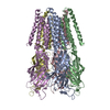
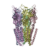
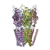
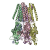
- PDB-5mur: X-ray structure of the F14'A mutant of GLIC in complex with propofol -
+ Open data

- Basic information
| Entry | Database: PDB / ID: 5mur | ||||||
|---|---|---|---|---|---|---|---|
| Title | X-ray structure of the F14'A mutant of GLIC in complex with propofol | ||||||
 Components Components | Proton-gated ion channel | ||||||
 Keywords Keywords | TRANSPORT PROTEIN / MEMBRANE PROTEIN | ||||||
| Function / homology |  Function and homology information Function and homology informationsodium channel activity / potassium channel activity / extracellular ligand-gated monoatomic ion channel activity / transmembrane signaling receptor activity / identical protein binding / plasma membrane Similarity search - Function | ||||||
| Biological species |  Gloeobacter violaceus (bacteria) Gloeobacter violaceus (bacteria) | ||||||
| Method |  X-RAY DIFFRACTION / SYNCHROTRON / MOLECULAR REPLACEMENT / Resolution: 3.1 Å X-RAY DIFFRACTION / SYNCHROTRON / MOLECULAR REPLACEMENT / Resolution: 3.1 Å | ||||||
 Authors Authors | Sauguet, L. / Fourati, Z. / Delarue, M. | ||||||
 Citation Citation | Journal: Cell Rep / Year: 2018 Title: Structural Basis for a Bimodal Allosteric Mechanism of General Anesthetic Modulation in Pentameric Ligand-Gated Ion Channels. Authors: Fourati, Z. / Howard, R.J. / Heusser, S.A. / Hu, H. / Ruza, R.R. / Sauguet, L. / Lindahl, E. / Delarue, M. | ||||||
| History |
|
- Structure visualization
| Structure viewer | Molecule: MolmilJmol/JSmol |
|---|
- Downloads & links
-Download
| PDBx/mmCIF format | 5mur.cif.gz | 652.8 KB | Display | PDBx/mmCIF format |
|---|---|---|---|---|
| PDB format | pdb5mur.ent.gz | 549.2 KB | Display | PDB format |
| PDBx/mmJSON format | 5mur.json.gz | Tree view | PDBx/mmJSON format | |
| Others |  Other downloads Other downloads |
-Validation report
| Arichive directory | https://data.pdbj.org/pub/pdb/validation_reports/mu/5murftp://data.pdbj.org/pub/pdb/validation_reports/mu/5mur | HTTPS FTP |
|---|
-Related structure data
| Related structure data |  5muoC  5mvnC  5mzqC  5nkjC  6emxC  4hfbS S: Starting model for refinement C: citing same article ( |
|---|---|
| Similar structure data |
-Links
 PDBj
PDBj


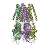
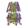
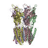
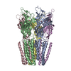
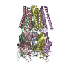
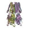
- Assembly
| Deposited unit | 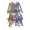
| ||||||||
|---|---|---|---|---|---|---|---|---|---|
| 1 |
| ||||||||
| Unit cell |
|
-Components
-Protein , 1 types, 5 molecules ABCDE
| #1: Protein | Mass: 36201.625 Da / Num. of mol.: 5 / Fragment: UNP residues 43-359 Source method: isolated from a genetically manipulated source Source: (gene. exp.) Gloeobacter violaceus (strain PCC 7421) (bacteria)Gene: glvI, glr4197 / Production host: Gloeobacter (bacteria) / References: UniProt: Q7NDN8 |
|---|
-Non-polymers , 6 types, 150 molecules 


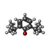

| #2: Chemical | ChemComp-ACT /  Mass: 59.044 Da / Num. of mol.: 10 / Source method: obtained synthetically / Formula: C2H3O2 Mass: 59.044 Da / Num. of mol.: 10 / Source method: obtained synthetically / Formula: C2H3O2#3: Chemical | ChemComp-CL /  Mass: 35.453 Da / Num. of mol.: 5 / Source method: obtained synthetically / Formula: Cl Mass: 35.453 Da / Num. of mol.: 5 / Source method: obtained synthetically / Formula: Cl#4: Chemical | ChemComp-NA /  Mass: 22.990 Da / Num. of mol.: 6 / Source method: obtained synthetically / Formula: Na Mass: 22.990 Da / Num. of mol.: 6 / Source method: obtained synthetically / Formula: Na#5: Chemical | ChemComp-PLC /  Mass: 622.834 Da / Num. of mol.: 5 / Source method: obtained synthetically / Formula: C32H65NO8P / Comment: phospholipid*YM Mass: 622.834 Da / Num. of mol.: 5 / Source method: obtained synthetically / Formula: C32H65NO8P / Comment: phospholipid*YM#6: Chemical |  Mass: 178.271 Da / Num. of mol.: 2 / Source method: obtained synthetically / Formula: C12H18O Mass: 178.271 Da / Num. of mol.: 2 / Source method: obtained synthetically / Formula: C12H18O#7: Water | ChemComp-HOH / | Mass: 18.015 Da / Num. of mol.: 122 / Source method: isolated from a natural source / Formula: H2O |
|---|
-Experimental details
-Experiment
| Experiment | Method: X-RAY DIFFRACTION / Number of used crystals: 1 |
|---|
- Sample preparation
| Crystal | Density Matthews: 5.15 Å3/Da / Density % sol: 76.1 % |
|---|---|
| Crystal grow | Temperature: 293 K / Method: vapor diffusion, hanging drop Details: 100 mM Na acetate pH4 12-15% PEG4K 200 mM NaSCN 16% glycerol 2% DMSO |
-Data collection
| Diffraction | Mean temperature: 80 K |
|---|---|
| Diffraction source | Source: SYNCHROTRON / Site: SOLEIL  / Beamline: PROXIMA 1 / Wavelength: 0.97 Å / Beamline: PROXIMA 1 / Wavelength: 0.97 Å |
| Detector | Type: DECTRIS PILATUS 6M-F / Detector: PIXEL / Date: Nov 30, 2011 |
| Radiation | Protocol: SINGLE WAVELENGTH / Monochromatic (M) / Laue (L): M / Scattering type: x-ray |
| Radiation wavelength | Wavelength: 0.97 Å / Relative weight: 1 |
| Reflection | Resolution: 3.1→49 Å / Num. obs: 66286 / % possible obs: 99 % / Redundancy: 3.5 % / Biso Wilson estimate: 94.16 Å2 / Net I/σ(I): 16.6 |
- Processing
| Software |
| ||||||||||||||||||||||||||||||||||||||||||||||||||||||||||||||||||||||||||||||||||||||||||||||||||||||||||||||||||||||||||||||||||||||||||||||||||||||
|---|---|---|---|---|---|---|---|---|---|---|---|---|---|---|---|---|---|---|---|---|---|---|---|---|---|---|---|---|---|---|---|---|---|---|---|---|---|---|---|---|---|---|---|---|---|---|---|---|---|---|---|---|---|---|---|---|---|---|---|---|---|---|---|---|---|---|---|---|---|---|---|---|---|---|---|---|---|---|---|---|---|---|---|---|---|---|---|---|---|---|---|---|---|---|---|---|---|---|---|---|---|---|---|---|---|---|---|---|---|---|---|---|---|---|---|---|---|---|---|---|---|---|---|---|---|---|---|---|---|---|---|---|---|---|---|---|---|---|---|---|---|---|---|---|---|---|---|---|---|---|---|
| Refinement | Method to determine structure: MOLECULAR REPLACEMENT Starting model: 4HFB Resolution: 3.1→20 Å / Cor.coef. Fo:Fc: 0.9175 / Cor.coef. Fo:Fc free: 0.9159 / SU R Cruickshank DPI: 0.599 / Cross valid method: THROUGHOUT / σ(F): 0 / SU R Blow DPI: 0.525 / SU Rfree Blow DPI: 0.272 / SU Rfree Cruickshank DPI: 0.284
| ||||||||||||||||||||||||||||||||||||||||||||||||||||||||||||||||||||||||||||||||||||||||||||||||||||||||||||||||||||||||||||||||||||||||||||||||||||||
| Displacement parameters | Biso mean: 101.8 Å2
| ||||||||||||||||||||||||||||||||||||||||||||||||||||||||||||||||||||||||||||||||||||||||||||||||||||||||||||||||||||||||||||||||||||||||||||||||||||||
| Refine analyze | Luzzati coordinate error obs: 0.439 Å | ||||||||||||||||||||||||||||||||||||||||||||||||||||||||||||||||||||||||||||||||||||||||||||||||||||||||||||||||||||||||||||||||||||||||||||||||||||||
| Refinement step | Cycle: 1 / Resolution: 3.1→20 Å
| ||||||||||||||||||||||||||||||||||||||||||||||||||||||||||||||||||||||||||||||||||||||||||||||||||||||||||||||||||||||||||||||||||||||||||||||||||||||
| Refine LS restraints |
| ||||||||||||||||||||||||||||||||||||||||||||||||||||||||||||||||||||||||||||||||||||||||||||||||||||||||||||||||||||||||||||||||||||||||||||||||||||||
| LS refinement shell | Resolution: 3.1→3.18 Å / Total num. of bins used: 20
| ||||||||||||||||||||||||||||||||||||||||||||||||||||||||||||||||||||||||||||||||||||||||||||||||||||||||||||||||||||||||||||||||||||||||||||||||||||||
| Refinement TLS params. | Method: refined / Refine-ID: X-RAY DIFFRACTION
| ||||||||||||||||||||||||||||||||||||||||||||||||||||||||||||||||||||||||||||||||||||||||||||||||||||||||||||||||||||||||||||||||||||||||||||||||||||||
| Refinement TLS group |
|
