 Movie
Movie Controller

[English] 日本語
 Yorodumi
- PDB-5jqq: Crystal structure of glucosyl-3-phosphoglycerate synthase from My... -
+ Open data

- Basic information
| Entry | Database: PDB / ID: 5jqq | ||||||
|---|---|---|---|---|---|---|---|
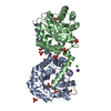
| Title | Crystal structure of glucosyl-3-phosphoglycerate synthase from Mycobacterium tuberculosis - apo form | ||||||
 Components Components | Glucosyl-3-phosphoglycerate synthase | ||||||
 Keywords Keywords | TRANSFERASE | ||||||
| Function / homology |  Function and homology informationglucosyl-3-phosphoglycerate synthase / UDP-glucose metabolic process / hexosyltransferase activity / magnesium ion binding / protein homodimerization activity Function and homology informationglucosyl-3-phosphoglycerate synthase / UDP-glucose metabolic process / hexosyltransferase activity / magnesium ion binding / protein homodimerization activitySimilarity search - Function | ||||||
| Biological species |  Mycobacterium tuberculosis H37Ra (bacteria) Mycobacterium tuberculosis H37Ra (bacteria) | ||||||
| Method | X-RAY DIFFRACTION / SYNCHROTRON / MOLECULAR REPLACEMENT / Resolution: 2.6 Å | ||||||
 Authors Authors | Albesa-Jove, D. / Urresti, S. / Gest, P.M. / van der Woerd, M. / Jackson, M. / Guerin, M.E. | ||||||
 Citation Citation | Journal: To Be Published Title: Crystal structure of glucosyl-3-phosphoglycerate synthase from Mycobacterium tuberculosis - apo form Authors: Albesa-Jove, D. / Romero-Garcia, J. / Sancho-Vaello, E. / Contreras, F.-X. / Rodrigo-Unzueta, A. / Comino, N. / Carreras-Gonzalez, A. / Arrasate, P. / Urresti, S. / Biarnes, X. / Planas, A. / Guerin, M.E. | ||||||
| History |
|
- Structure visualization
| Structure viewer | Molecule: MolmilJmol/JSmol |
|---|
- Downloads & links
-Download
| PDBx/mmCIF format | 5jqq.cif.gz | 71.1 KB | Display | PDBx/mmCIF format |
|---|---|---|---|---|
| PDB format | pdb5jqq.ent.gz | 50.9 KB | Display | PDB format |
| PDBx/mmJSON format | 5jqq.json.gz | Tree view | PDBx/mmJSON format | |
| Others |  Other downloads Other downloads |
-Validation report
| Arichive directory | https://data.pdbj.org/pub/pdb/validation_reports/jq/5jqqftp://data.pdbj.org/pub/pdb/validation_reports/jq/5jqq | HTTPS FTP |
|---|
-Related structure data
| Related structure data |  1ckjS S: Starting model for refinement |
|---|---|
| Similar structure data |


-Links
 PDBj
PDBj

- Assembly
| Deposited unit | 
| ||||||||
|---|---|---|---|---|---|---|---|---|---|
| 1 | 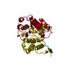
| ||||||||
| Unit cell |
| ||||||||
| Components on special symmetry positions |
|
-Components
| #1: Protein | Mass: 36582.656 Da / Num. of mol.: 1 Source method: isolated from a genetically manipulated source Source: (gene. exp.) Mycobacterium tuberculosis H37Ra (bacteria)Gene: gpgS, Rv1208 / Production host: Escherichia coli (E. coli)References: UniProt: P9WMW9, glucosyl-3-phosphoglycerate synthase | ||
|---|---|---|---|
| #2: Chemical | Glycerol  Mass: 92.094 Da / Num. of mol.: 2 / Source method: obtained synthetically / Formula: C3H8O3 Mass: 92.094 Da / Num. of mol.: 2 / Source method: obtained synthetically / Formula: C3H8O3#3: Water | ChemComp-HOH / | Water Mass: 18.015 Da / Num. of mol.: 59 / Source method: isolated from a natural source / Formula: H2O Mass: 18.015 Da / Num. of mol.: 59 / Source method: isolated from a natural source / Formula: H2O |
-Experimental details
-Experiment
| Experiment | Method: X-RAY DIFFRACTION / Number of used crystals: 1 |
|---|
- Sample preparation
| Crystal | Density Matthews: 4.26 Å3/Da / Density % sol: 71.14 % |
|---|---|
| Crystal grow | Temperature: 289 K / Method: vapor diffusion, hanging drop / pH: 6.5 Details: 1 mM MgCl2, 100 mM sodium cacodylate pH 6.5, 200 mM trisodium citrate, 30%(v/v) 2-propanol, VAPOR DIFFUSION, HANGING DROP, temperature 289K |
-Data collection
| Diffraction | Mean temperature: 100 K |
|---|---|
| Diffraction source | Source: SYNCHROTRON / Site: ALS  / Beamline: 4.2.2 / Wavelength: 1 Å / Beamline: 4.2.2 / Wavelength: 1 Å |
| Detector | Type: NOIR-1 / Detector: CCD / Date: Jul 22, 2008 Details: Rosenbaum-Rock Si(111) sagitally focused monochromator |
| Radiation | Protocol: SINGLE WAVELENGTH / Monochromatic (M) / Laue (L): M / Scattering type: x-ray |
| Radiation wavelength | Wavelength: 1 Å / Relative weight: 1 |
| Reflection | Resolution: 2.6→41.77 Å / Num. obs: 37009 / % possible obs: 100 % / Redundancy: 7 % / Rsym value: 0.106 / Net I/σ(I): 9.1 |
| Reflection shell | Resolution: 2.6→2.69 Å / Redundancy: 6.5 % / Rmerge(I) obs: 0.48 / Mean I/σ(I) obs: 3 / % possible all: 100 |
- Processing
| Software |
| |||||||||||||||||||||||||||||||||||||||||||||||||||||||||||||||||||||||||||||||||||||||||||||||||||||||||
|---|---|---|---|---|---|---|---|---|---|---|---|---|---|---|---|---|---|---|---|---|---|---|---|---|---|---|---|---|---|---|---|---|---|---|---|---|---|---|---|---|---|---|---|---|---|---|---|---|---|---|---|---|---|---|---|---|---|---|---|---|---|---|---|---|---|---|---|---|---|---|---|---|---|---|---|---|---|---|---|---|---|---|---|---|---|---|---|---|---|---|---|---|---|---|---|---|---|---|---|---|---|---|---|---|---|---|
| Refinement | Method to determine structure: MOLECULAR REPLACEMENT Starting model: 1CKJ Resolution: 2.6→39.077 Å / SU ML: 0.32 / Cross valid method: FREE R-VALUE / σ(F): 1.16 / Phase error: 26.54
| |||||||||||||||||||||||||||||||||||||||||||||||||||||||||||||||||||||||||||||||||||||||||||||||||||||||||
| Solvent computation | Shrinkage radii: 0.9 Å / VDW probe radii: 1.2 Å | |||||||||||||||||||||||||||||||||||||||||||||||||||||||||||||||||||||||||||||||||||||||||||||||||||||||||
| Refinement step | Cycle: LAST / Resolution: 2.6→39.077 Å
| |||||||||||||||||||||||||||||||||||||||||||||||||||||||||||||||||||||||||||||||||||||||||||||||||||||||||
| Refine LS restraints |
| |||||||||||||||||||||||||||||||||||||||||||||||||||||||||||||||||||||||||||||||||||||||||||||||||||||||||
| LS refinement shell |
|
