 Movie
Movie Controller


+ Open data

- Basic information
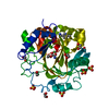
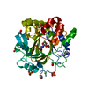
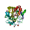
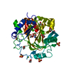
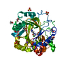
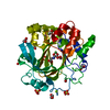
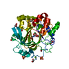
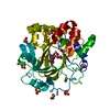
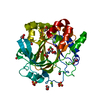
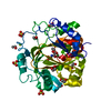
| Entry | Database: PDB / ID: 5f5c | ||||||
|---|---|---|---|---|---|---|---|
| Title | Crystal Structure of human JMJD2D complexed with KDOPP7 | ||||||
 Components Components | Lysine-specific demethylase 4D | ||||||
 Keywords Keywords |  OXIDOREDUCTASE / DOUBLE-STRANDED BETA HELIX / DEMETHYLASE / OXYGENASE / Structural Genomics / Structural Genomics Consortium / SGC OXIDOREDUCTASE / DOUBLE-STRANDED BETA HELIX / DEMETHYLASE / OXYGENASE / Structural Genomics / Structural Genomics Consortium / SGC | ||||||
| Function / homology |  Function and homology information Function and homology informationpositive regulation of chromatin binding / histone H3K9me2/H3K9me3 demethylase activity / [histone H3]-trimethyl-L-lysine9 demethylase / positive regulation of double-strand break repair via nonhomologous end joining / histone H3K9 demethylase activity / histone demethylase activity / pericentric heterochromatin / cellular response to ionizing radiation / double-strand break repair via homologous recombination / regulation of protein phosphorylation ...positive regulation of chromatin binding / histone H3K9me2/H3K9me3 demethylase activity / [histone H3]-trimethyl-L-lysine9 demethylase / positive regulation of double-strand break repair via nonhomologous end joining / histone H3K9 demethylase activity / histone demethylase activity / pericentric heterochromatin / cellular response to ionizing radiation / double-strand break repair via homologous recombination / regulation of protein phosphorylation / HDMs demethylate histones / chromatin DNA binding / site of double-strand break / regulation of gene expression / blood microparticle / damaged DNA binding / chromatin remodeling / inflammatory response / chromatin / nucleoplasm / metal ion binding / nucleusSimilarity search - Function | ||||||
| Biological species |  Homo sapiens (human) Homo sapiens (human) | ||||||
| Method | X-RAY DIFFRACTION / MOLECULAR REPLACEMENT / molecular replacement / Resolution: 1.88 Å | ||||||
 Authors Authors | Krojer, T. / Vollmar, M. / Crawley, L. / Bradley, A.R. / Szykowska, A. / Ruda, G.F. / Yang, H. / Burgess-Brown, N. / Brennan, P. / Bountra, C. ...Krojer, T. / Vollmar, M. / Crawley, L. / Bradley, A.R. / Szykowska, A. / Ruda, G.F. / Yang, H. / Burgess-Brown, N. / Brennan, P. / Bountra, C. / Arrowsmith, C.H. / Edwards, A. / Oppermann, U. / von Delft, F. / Structural Genomics Consortium (SGC) | ||||||
 Citation Citation | Journal: J.Med.Chem. / Year: 2016 Title: 8-Substituted Pyrido[3,4-d]pyrimidin-4(3H)-one Derivatives As Potent, Cell Permeable, KDM4 (JMJD2) and KDM5 (JARID1) Histone Lysine Demethylase Inhibitors. Authors: Bavetsias, V. / Lanigan, R.M. / Ruda, G.F. / Atrash, B. / McLaughlin, M.G. / Tumber, A. / Mok, N.Y. / Le Bihan, Y.V. / Dempster, S. / Boxall, K.J. / Jeganathan, F. / Hatch, S.B. / Savitsky, ...Authors: Bavetsias, V. / Lanigan, R.M. / Ruda, G.F. / Atrash, B. / McLaughlin, M.G. / Tumber, A. / Mok, N.Y. / Le Bihan, Y.V. / Dempster, S. / Boxall, K.J. / Jeganathan, F. / Hatch, S.B. / Savitsky, P. / Velupillai, S. / Krojer, T. / England, K.S. / Sejberg, J. / Thai, C. / Donovan, A. / Pal, A. / Scozzafava, G. / Bennett, J.M. / Kawamura, A. / Johansson, C. / Szykowska, A. / Gileadi, C. / Burgess-Brown, N.A. / von Delft, F. / Oppermann, U. / Walters, Z. / Shipley, J. / Raynaud, F.I. / Westaway, S.M. / Prinjha, R.K. / Fedorov, O. / Burke, R. / Schofield, C.J. / Westwood, I.M. / Bountra, C. / Muller, S. / van Montfort, R.L. / Brennan, P.E. / Blagg, J. | ||||||
| History |
|
- Structure visualization
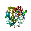
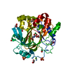
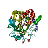
| Structure viewer | Molecule: MolmilJmol/JSmol |
|---|
- Downloads & links
-Download
| PDBx/mmCIF format | 5f5c.cif.gz | 164.2 KB | Display | PDBx/mmCIF format |
|---|---|---|---|---|
| PDB format | pdb5f5c.ent.gz | 127.9 KB | Display | PDB format |
| PDBx/mmJSON format | 5f5c.json.gz | Tree view | PDBx/mmJSON format | |
| Others |  Other downloads Other downloads |
-Validation report
| Arichive directory | https://data.pdbj.org/pub/pdb/validation_reports/f5/5f5cftp://data.pdbj.org/pub/pdb/validation_reports/f5/5f5c | HTTPS FTP |
|---|
-Related structure data
| Related structure data | 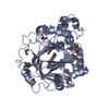 5f2sC 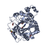 5f2wC 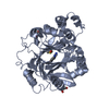 5f32C 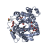 5f37C 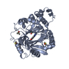 5f39C 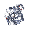 5f3cC 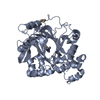 5f3eC 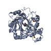 5f3gC 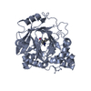 5f3iC 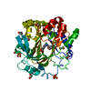 5f5aC 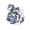 5f5iC  5fplC 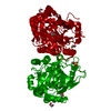 4d6qS S: Starting model for refinement C: citing same article ( |
|---|---|
| Similar structure data |


-Links
 PDBj
PDBj
- Assembly
| Deposited unit | 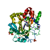
| ||||||||
|---|---|---|---|---|---|---|---|---|---|
| 1 |
| ||||||||
| Unit cell |
| ||||||||
| Components on special symmetry positions |
|
-Components
-Protein , 1 types, 1 molecules A
| #1: Protein | Mass: 40017.348 Da / Num. of mol.: 1 / Fragment: Oxidoreductase-2OG Source method: isolated from a genetically manipulated source Source: (gene. exp.) Homo sapiens (human) / Gene: KDM4D, JHDM3D, JMJD2D / Plasmid: pNIC28-Bsa4 / Production host:  Escherichia coli (E. coli) Escherichia coli (E. coli)References: UniProt: Q6B0I6, Oxidoreductases; Acting on paired donors, with incorporation or reduction of molecular oxygen; With 2-oxoglutarate as one donor, and incorporation of one atom of oxygen into each donor |
|---|
-Non-polymers , 6 types, 422 molecules 


| #2: Chemical | ChemComp-ZN /  Mass: 65.409 Da / Num. of mol.: 1 / Source method: obtained synthetically / Formula: Zn Mass: 65.409 Da / Num. of mol.: 1 / Source method: obtained synthetically / Formula: Zn | ||||||
|---|---|---|---|---|---|---|---|
| #3: Chemical | ChemComp-NI / Nickel Mass: 58.693 Da / Num. of mol.: 1 / Source method: obtained synthetically / Formula: Ni Mass: 58.693 Da / Num. of mol.: 1 / Source method: obtained synthetically / Formula: Ni | ||||||
| #4: Chemical | ChemComp-EDO / Ethylene glycol Mass: 62.068 Da / Num. of mol.: 5 / Source method: obtained synthetically / Formula: C2H6O2 Mass: 62.068 Da / Num. of mol.: 5 / Source method: obtained synthetically / Formula: C2H6O2#5: Chemical | Sulfate Mass: 96.063 Da / Num. of mol.: 3 / Source method: obtained synthetically / Formula: SO4 Mass: 96.063 Da / Num. of mol.: 3 / Source method: obtained synthetically / Formula: SO4#6: Chemical | ChemComp-5V3 / |  Mass: 266.298 Da / Num. of mol.: 1 / Source method: obtained synthetically / Formula: C15H14N4O Mass: 266.298 Da / Num. of mol.: 1 / Source method: obtained synthetically / Formula: C15H14N4O#7: Water | ChemComp-HOH / | WaterMass: 18.015 Da / Num. of mol.: 411 / Source method: isolated from a natural source / Formula: H2O |
-Experimental details
-Experiment
| Experiment | Method: X-RAY DIFFRACTION / Number of used crystals: 1 |
|---|
- Sample preparation
| Crystal | Density Matthews: 2.41 Å3/Da / Density % sol: 49.05 % |
|---|---|
| Crystal grow | Temperature: 293 K / Method: vapor diffusion, sitting drop / pH: 6.8 Details: 26% PEG3350 , 0.1M HEPES pH 6.8 , 0.2M ammonium sulfate |
-Data collection
| Diffraction | Mean temperature: 100 K |
|---|---|
| Diffraction source | Source: ROTATING ANODE / Type: BRUKER AXS MICROSTAR / Wavelength: 1.5418 Å |
| Detector | Type: Bruker Platinum 135 / Detector: CCD / Date: Apr 28, 2014 |
| Radiation | Protocol: SINGLE WAVELENGTH / Monochromatic (M) / Laue (L): M / Scattering type: x-ray |
| Radiation wavelength | Wavelength: 1.5418 Å / Relative weight: 1 |
| Reflection | Resolution: 1.88→64.68 Å / Num. all: 32698 / Num. obs: 32698 / % possible obs: 99.8 % / Observed criterion σ(F): -3 / Observed criterion σ(I): -3 / Redundancy: 10.4 % / Rmerge(I) obs: 0.129 / Net I/σ(I): 13.5 |
| Reflection shell | Resolution: 1.88→1.92 Å / Redundancy: 3.95 % / Rmerge(I) obs: 0.518 / Mean I/σ(I) obs: 1.96 / % possible all: 97.5 |
-Phasing
| Phasing | Method: molecular replacement |
|---|
- Processing
| Software |
| ||||||||||||||||||||||||||||||||||||||||||||||||||||||||||||||||||||||||||||||||||||||||||||||||||||||||||||||||||||||||||||||||||||||||||||||||||||||||||||||||||||||||||||||||||||||||||||||||||||||||
|---|---|---|---|---|---|---|---|---|---|---|---|---|---|---|---|---|---|---|---|---|---|---|---|---|---|---|---|---|---|---|---|---|---|---|---|---|---|---|---|---|---|---|---|---|---|---|---|---|---|---|---|---|---|---|---|---|---|---|---|---|---|---|---|---|---|---|---|---|---|---|---|---|---|---|---|---|---|---|---|---|---|---|---|---|---|---|---|---|---|---|---|---|---|---|---|---|---|---|---|---|---|---|---|---|---|---|---|---|---|---|---|---|---|---|---|---|---|---|---|---|---|---|---|---|---|---|---|---|---|---|---|---|---|---|---|---|---|---|---|---|---|---|---|---|---|---|---|---|---|---|---|---|---|---|---|---|---|---|---|---|---|---|---|---|---|---|---|---|---|---|---|---|---|---|---|---|---|---|---|---|---|---|---|---|---|---|---|---|---|---|---|---|---|---|---|---|---|---|---|---|---|
| Refinement | Method to determine structure: MOLECULAR REPLACEMENT Starting model: 4d6q Resolution: 1.88→51.92 Å / Cor.coef. Fo:Fc: 0.95 / Cor.coef. Fo:Fc free: 0.925 / SU B: 5.898 / SU ML: 0.092 / Cross valid method: THROUGHOUT / σ(F): 0 / ESU R: 0.142 / ESU R Free: 0.132 / Stereochemistry target values: MAXIMUM LIKELIHOOD Details: HYDROGENS HAVE BEEN ADDED IN THE RIDING POSITIONS U VALUES : WITH TLS ADDED
| ||||||||||||||||||||||||||||||||||||||||||||||||||||||||||||||||||||||||||||||||||||||||||||||||||||||||||||||||||||||||||||||||||||||||||||||||||||||||||||||||||||||||||||||||||||||||||||||||||||||||
| Solvent computation | Ion probe radii: 0.8 Å / Shrinkage radii: 0.8 Å / VDW probe radii: 1.2 Å / Solvent model: MASK | ||||||||||||||||||||||||||||||||||||||||||||||||||||||||||||||||||||||||||||||||||||||||||||||||||||||||||||||||||||||||||||||||||||||||||||||||||||||||||||||||||||||||||||||||||||||||||||||||||||||||
| Displacement parameters | Biso max: 61.85 Å2 / Biso mean: 13.863 Å2 / Biso min: 2.78 Å2
| ||||||||||||||||||||||||||||||||||||||||||||||||||||||||||||||||||||||||||||||||||||||||||||||||||||||||||||||||||||||||||||||||||||||||||||||||||||||||||||||||||||||||||||||||||||||||||||||||||||||||
| Refinement step | Cycle: final / Resolution: 1.88→51.92 Å
| ||||||||||||||||||||||||||||||||||||||||||||||||||||||||||||||||||||||||||||||||||||||||||||||||||||||||||||||||||||||||||||||||||||||||||||||||||||||||||||||||||||||||||||||||||||||||||||||||||||||||
| Refine LS restraints |
| ||||||||||||||||||||||||||||||||||||||||||||||||||||||||||||||||||||||||||||||||||||||||||||||||||||||||||||||||||||||||||||||||||||||||||||||||||||||||||||||||||||||||||||||||||||||||||||||||||||||||
| LS refinement shell | Resolution: 1.88→1.929 Å / Total num. of bins used: 20
| ||||||||||||||||||||||||||||||||||||||||||||||||||||||||||||||||||||||||||||||||||||||||||||||||||||||||||||||||||||||||||||||||||||||||||||||||||||||||||||||||||||||||||||||||||||||||||||||||||||||||
| Refinement TLS params. | Method: refined / Refine-ID: X-RAY DIFFRACTION
| ||||||||||||||||||||||||||||||||||||||||||||||||||||||||||||||||||||||||||||||||||||||||||||||||||||||||||||||||||||||||||||||||||||||||||||||||||||||||||||||||||||||||||||||||||||||||||||||||||||||||
| Refinement TLS group |
|
