 Movie
Movie Controller


+ Open data

- Basic information
| Entry | Database: PDB / ID: 5fpl | ||||||
|---|---|---|---|---|---|---|---|
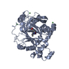
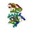
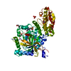
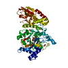
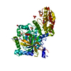
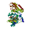
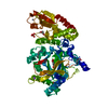
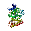
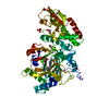
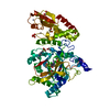
| Title | Crystal structure of human JARID1B in complex with CCT363901 | ||||||
 Components Components | LYSINE-SPECIFIC DEMETHYLASE 5B, LYSINE-SPECIFIC DEMETHYLASE 5B | ||||||
 Keywords Keywords | OXIDOREDUCTASE / LYSINE-SPECIFIC DEMETHYLASE 5B | ||||||
| Function / homology |  Function and homology information Function and homology informationregulation of estradiol secretion / mammary duct terminal end bud growth / uterus morphogenesis / TFAP2 (AP-2) family regulates transcription of cell cycle factors / positive regulation of mammary gland epithelial cell proliferation / [histone H3]-trimethyl-L-lysine4 demethylase / histone H3K4me/H3K4me2/H3K4me3 demethylase activity / lens fiber cell differentiation / histone H3K4 demethylase activity / branching involved in mammary gland duct morphogenesis ...regulation of estradiol secretion / mammary duct terminal end bud growth / uterus morphogenesis / TFAP2 (AP-2) family regulates transcription of cell cycle factors / positive regulation of mammary gland epithelial cell proliferation / [histone H3]-trimethyl-L-lysine4 demethylase / histone H3K4me/H3K4me2/H3K4me3 demethylase activity / lens fiber cell differentiation / histone H3K4 demethylase activity / branching involved in mammary gland duct morphogenesis / histone demethylase activity / single fertilization / response to fungicide / cellular response to fibroblast growth factor stimulus / cellular response to leukemia inhibitory factor / Chromatin modifications during the maternal to zygotic transition (MZT) / post-embryonic development / HDMs demethylate histones / sequence-specific double-stranded DNA binding / transcription corepressor activity / rhythmic process / histone binding / nucleic acid binding / chromatin remodeling / negative regulation of DNA-templated transcription / positive regulation of gene expression / regulation of DNA-templated transcription / chromatin / DNA binding / zinc ion binding / nucleoplasm / nucleus / cytosol Similarity search - Function | ||||||
| Biological species |  HOMO SAPIENS (human) HOMO SAPIENS (human) | ||||||
| Method |  X-RAY DIFFRACTION / SYNCHROTRON / MOLECULAR REPLACEMENT / Resolution: 2.35 Å X-RAY DIFFRACTION / SYNCHROTRON / MOLECULAR REPLACEMENT / Resolution: 2.35 Å | ||||||
 Authors Authors | Srikannathasan, V. / Yann-Vai, L.B. / Nowak, R. / Johansson, C. / Gileadi, C. / von Delft, F. / Arrowsmith, C.H. / Bountra, C. / Edwards, A. / Brennan, P. ...Srikannathasan, V. / Yann-Vai, L.B. / Nowak, R. / Johansson, C. / Gileadi, C. / von Delft, F. / Arrowsmith, C.H. / Bountra, C. / Edwards, A. / Brennan, P. / Huber, K. / Oppermann, U. | ||||||
 Citation Citation | Journal: J.Med.Chem. / Year: 2016 Title: 8-Substituted Pyrido[3,4-D]Pyrimidin-4(3H)-One Derivatives as Potent, Cell Permeable, Kdm4 (Jmjd2) and Kdm5 (Jarid1) Histone Lysine Demethylase Inhibitors. Authors: Bavetsias, V. / Lanigan, R.M. / Ruda, G.F. / Atrash, B. / Mclaughlin, M.G. / Tumber, A. / Mok, N.Y. / Le Bihan, Y. / Dempster, S. / Boxall, K.J. / Jeganathan, F. / Hatch, S.B. / Savitsky, P. ...Authors: Bavetsias, V. / Lanigan, R.M. / Ruda, G.F. / Atrash, B. / Mclaughlin, M.G. / Tumber, A. / Mok, N.Y. / Le Bihan, Y. / Dempster, S. / Boxall, K.J. / Jeganathan, F. / Hatch, S.B. / Savitsky, P. / Velupillai, S. / Krojer, T. / England, K.S. / Sejberg, J. / Thai, C. / Donovan, A. / Pal, A. / Scozzafava, G. / Bennett, J.M. / Kawamura, A. / Johansson, C. / Szykowska, A. / Gileadi, C. / Burgess-Brown, N.A. / von Delft, F. / Oppermann, U. / Walters, Z. / Shipley, J. / Raynaud, F.I. / Westaway, S.M. / Prinjha, R.K. / Fedorov, O. / Burke, R. / Schofield, C.J. / Westwood, I.M. / Bountra, C. / Muller, S. / Van Montfort, R.L.M. / Brennan, P.E. / Blagg, J. | ||||||
| History |
|
- Structure visualization
| Structure viewer | Molecule: MolmilJmol/JSmol |
|---|
- Downloads & links
-Download
| PDBx/mmCIF format | 5fpl.cif.gz | 211.9 KB | Display | PDBx/mmCIF format |
|---|---|---|---|---|
| PDB format | pdb5fpl.ent.gz | 169.5 KB | Display | PDB format |
| PDBx/mmJSON format | 5fpl.json.gz | Tree view | PDBx/mmJSON format | |
| Others |  Other downloads Other downloads |
-Validation report
| Arichive directory | https://data.pdbj.org/pub/pdb/validation_reports/fp/5fplftp://data.pdbj.org/pub/pdb/validation_reports/fp/5fpl | HTTPS FTP |
|---|
-Related structure data
| Related structure data | 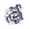 5f2sC 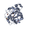 5f2wC 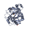 5f32C  5f37C 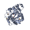 5f39C 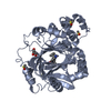 5f3cC 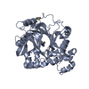 5f3eC 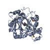 5f3gC 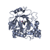 5f3iC 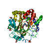 5f5aC 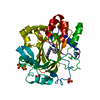 5f5cC 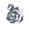 5f5iC 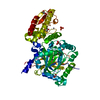 5a1fS S: Starting model for refinement C: citing same article ( |
|---|---|
| Similar structure data |


-Links
 PDBj
PDBj

- Assembly
| Deposited unit | 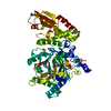
| ||||||||
|---|---|---|---|---|---|---|---|---|---|
| 1 |
| ||||||||
| Unit cell |
|
-Components
-Protein , 1 types, 1 molecules A
| #1: Protein | Mass: 55370.570 Da / Num. of mol.: 1 / Fragment: JMJC DOMAIN, RESIDUES 25-101,374-772 Source method: isolated from a genetically manipulated source Source: (gene. exp.) HOMO SAPIENS (human) / Plasmid: PFB-LIC-BSE / Cell line (production host): SF9 / Production host:   SPODOPTERA FRUGIPERDA (fall armyworm) / References: UniProt: Q9UGL1 SPODOPTERA FRUGIPERDA (fall armyworm) / References: UniProt: Q9UGL1 |
|---|
-Non-polymers , 6 types, 286 molecules 


| #2: Chemical | ChemComp-ZN /  Mass: 65.409 Da / Num. of mol.: 1 / Source method: obtained synthetically / Formula: Zn Mass: 65.409 Da / Num. of mol.: 1 / Source method: obtained synthetically / Formula: Zn | ||||||||
|---|---|---|---|---|---|---|---|---|---|
| #3: Chemical |  Mass: 54.938 Da / Num. of mol.: 2 / Source method: obtained synthetically / Formula: Mn Mass: 54.938 Da / Num. of mol.: 2 / Source method: obtained synthetically / Formula: Mn#4: Chemical | ChemComp-QAY / |  Mass: 256.263 Da / Num. of mol.: 1 / Source method: obtained synthetically / Formula: C12H12N6O Mass: 256.263 Da / Num. of mol.: 1 / Source method: obtained synthetically / Formula: C12H12N6O#5: Chemical |  Mass: 62.068 Da / Num. of mol.: 2 / Source method: obtained synthetically / Formula: C2H6O2 Mass: 62.068 Da / Num. of mol.: 2 / Source method: obtained synthetically / Formula: C2H6O2#6: Chemical | ChemComp-DMS / |  Mass: 78.133 Da / Num. of mol.: 1 / Source method: obtained synthetically / Formula: C2H6OS / Comment: DMSO, precipitant*YM Mass: 78.133 Da / Num. of mol.: 1 / Source method: obtained synthetically / Formula: C2H6OS / Comment: DMSO, precipitant*YM#7: Water | ChemComp-HOH / | Mass: 18.015 Da / Num. of mol.: 279 / Source method: isolated from a natural source / Formula: H2O |
-Details
| Has protein modification | Y |
|---|---|
| Sequence details | RESIDUE NUMBER 0-MET FROM THE CONSTRUCT. KDM5B START WITH PHE26. GGGG (101-105) FOUR GLYCINE LINKER ...RESIDUE NUMBER 0-MET FROM THE CONSTRUCT. KDM5B START WITH PHE26. GGGG (101-105) FOUR GLYCINE LINKER INTRODUCED |
-Experimental details
-Experiment
| Experiment | Method: X-RAY DIFFRACTION / Number of used crystals: 1 |
|---|
- Sample preparation
| Crystal | Density Matthews: 4.05 Å3/Da / Density % sol: 69.65 % / Description: NONE |
|---|---|
| Crystal grow | pH: 8 Details: 0.1M HEPES PH 8.0, 0.8M POTASSIUM PHOSPHATE-DIBASIC, 0.8M SODIUM PHOSPHATE MONOBASIC |
-Data collection
| Diffraction | Mean temperature: 100 K |
|---|---|
| Diffraction source | Source: SYNCHROTRON / Site: Diamond  / Beamline: I02 / Wavelength: 0.9795 / Beamline: I02 / Wavelength: 0.9795 |
| Detector | Type: DECTRIS PILATUS 6M / Detector: PIXEL / Date: Jul 22, 2015 / Details: MIRRORS |
| Radiation | Monochromator: M / Protocol: SINGLE WAVELENGTH / Monochromatic (M) / Laue (L): M / Scattering type: x-ray |
| Radiation wavelength | Wavelength: 0.9795 Å / Relative weight: 1 |
| Reflection | Resolution: 2.35→46.17 Å / Num. obs: 38565 / % possible obs: 99.7 % / Observed criterion σ(I): 2 / Redundancy: 20.1 % / Biso Wilson estimate: 65.42 Å2 / Rmerge(I) obs: 0.15 / Net I/σ(I): 14.27 |
| Reflection shell | Resolution: 2.35→2.43 Å / Redundancy: 19.7 % / Rmerge(I) obs: 0.15 / Mean I/σ(I) obs: 14.27 / % possible all: 99.6 |
- Processing
| Software |
| ||||||||||||||||||||||||||||||||||||||||||||||||||||||||||||||||||||||||||||||||||||||||||||||||||||||||||||||||||
|---|---|---|---|---|---|---|---|---|---|---|---|---|---|---|---|---|---|---|---|---|---|---|---|---|---|---|---|---|---|---|---|---|---|---|---|---|---|---|---|---|---|---|---|---|---|---|---|---|---|---|---|---|---|---|---|---|---|---|---|---|---|---|---|---|---|---|---|---|---|---|---|---|---|---|---|---|---|---|---|---|---|---|---|---|---|---|---|---|---|---|---|---|---|---|---|---|---|---|---|---|---|---|---|---|---|---|---|---|---|---|---|---|---|---|---|
| Refinement | Method to determine structure: MOLECULAR REPLACEMENT Starting model: PDB ENTRY 5A1F Resolution: 2.35→46.17 Å / Cor.coef. Fo:Fc: 0.9433 / Cor.coef. Fo:Fc free: 0.9383 / SU R Cruickshank DPI: 0.19 / Cross valid method: THROUGHOUT / σ(F): 0 / SU R Blow DPI: 0.203 / SU Rfree Blow DPI: 0.162 / SU Rfree Cruickshank DPI: 0.158
| ||||||||||||||||||||||||||||||||||||||||||||||||||||||||||||||||||||||||||||||||||||||||||||||||||||||||||||||||||
| Displacement parameters | Biso mean: 65.2 Å2
| ||||||||||||||||||||||||||||||||||||||||||||||||||||||||||||||||||||||||||||||||||||||||||||||||||||||||||||||||||
| Refine analyze | Luzzati coordinate error obs: 0.286 Å | ||||||||||||||||||||||||||||||||||||||||||||||||||||||||||||||||||||||||||||||||||||||||||||||||||||||||||||||||||
| Refinement step | Cycle: LAST / Resolution: 2.35→46.17 Å
| ||||||||||||||||||||||||||||||||||||||||||||||||||||||||||||||||||||||||||||||||||||||||||||||||||||||||||||||||||
| Refine LS restraints |
| ||||||||||||||||||||||||||||||||||||||||||||||||||||||||||||||||||||||||||||||||||||||||||||||||||||||||||||||||||
| LS refinement shell | Resolution: 2.35→2.41 Å / Total num. of bins used: 19
| ||||||||||||||||||||||||||||||||||||||||||||||||||||||||||||||||||||||||||||||||||||||||||||||||||||||||||||||||||
| Refinement TLS params. | Method: refined / Refine-ID: X-RAY DIFFRACTION
| ||||||||||||||||||||||||||||||||||||||||||||||||||||||||||||||||||||||||||||||||||||||||||||||||||||||||||||||||||
| Refinement TLS group |
|
