 Movie
Movie Controller

[English] 日本語
 Yorodumi
- PDB-5dne: Crystal structure of the Asn-bound guinea pig L-asparaginase 1 ca... -
+ Open data

- Basic information
| Entry | Database: PDB / ID: 5dne | ||||||
|---|---|---|---|---|---|---|---|
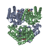
| Title | Crystal structure of the Asn-bound guinea pig L-asparaginase 1 catalytic domain active site mutant K188M | ||||||
 Components Components | L-asparaginase | ||||||
 Keywords Keywords | HYDROLASE / asparaginase | ||||||
| Function / homology |  Function and homology information Function and homology information | ||||||
| Biological species |  Cavia porcellus (domestic guinea pig) Cavia porcellus (domestic guinea pig) | ||||||
| Method |  X-RAY DIFFRACTION / SYNCHROTRON / MOLECULAR REPLACEMENT / Resolution: 2.39 Å X-RAY DIFFRACTION / SYNCHROTRON / MOLECULAR REPLACEMENT / Resolution: 2.39 Å | ||||||
 Authors Authors | Schalk, A.M. / Lavie, A. | ||||||
 Citation Citation | Journal: J.Biol.Chem. / Year: 2016 Title: Experimental Data in Support of a Direct Displacement Mechanism for Type I/II l-Asparaginases. Authors: Schalk, A.M. / Antansijevic, A. / Caffrey, M. / Lavie, A. | ||||||
| History |
|
- Structure visualization
| Structure viewer | Molecule: MolmilJmol/JSmol |
|---|
- Downloads & links
-Download
| PDBx/mmCIF format | 5dne.cif.gz | 296.4 KB | Display | PDBx/mmCIF format |
|---|---|---|---|---|
| PDB format | pdb5dne.ent.gz | 231.7 KB | Display | PDB format |
| PDBx/mmJSON format | 5dne.json.gz | Tree view | PDBx/mmJSON format | |
| Others |  Other downloads Other downloads |
-Validation report
| Arichive directory | https://data.pdbj.org/pub/pdb/validation_reports/dn/5dneftp://data.pdbj.org/pub/pdb/validation_reports/dn/5dne | HTTPS FTP |
|---|
-Related structure data
| Related structure data |  5dncC 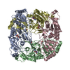 5dndC  4r8lS S: Starting model for refinement C: citing same article ( |
|---|---|
| Similar structure data |


-Links
 PDBj
PDBj- Assembly
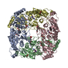
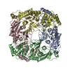
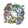
| Deposited unit | 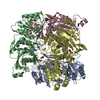
| ||||||||||||||||||||||||||||||||||||||||||||||||||||||||||||||||||||||||||||||||||||||||||||||||||||||||||||||||||||||||||||||||||||||||||||||||||||||
|---|---|---|---|---|---|---|---|---|---|---|---|---|---|---|---|---|---|---|---|---|---|---|---|---|---|---|---|---|---|---|---|---|---|---|---|---|---|---|---|---|---|---|---|---|---|---|---|---|---|---|---|---|---|---|---|---|---|---|---|---|---|---|---|---|---|---|---|---|---|---|---|---|---|---|---|---|---|---|---|---|---|---|---|---|---|---|---|---|---|---|---|---|---|---|---|---|---|---|---|---|---|---|---|---|---|---|---|---|---|---|---|---|---|---|---|---|---|---|---|---|---|---|---|---|---|---|---|---|---|---|---|---|---|---|---|---|---|---|---|---|---|---|---|---|---|---|---|---|---|---|---|
| 1 |
| ||||||||||||||||||||||||||||||||||||||||||||||||||||||||||||||||||||||||||||||||||||||||||||||||||||||||||||||||||||||||||||||||||||||||||||||||||||||
| Unit cell |
| ||||||||||||||||||||||||||||||||||||||||||||||||||||||||||||||||||||||||||||||||||||||||||||||||||||||||||||||||||||||||||||||||||||||||||||||||||||||
| Noncrystallographic symmetry (NCS) | NCS domain:
NCS domain segments: Component-ID: _ / Beg auth comp-ID: GLU / Beg label comp-ID: GLU / Refine code: _
NCS ensembles :
|
-Components
| #1: Protein | Mass: 63386.758 Da / Num. of mol.: 4 / Mutation: K188M Source method: isolated from a genetically manipulated source Details: The first 23 residues are from the tag hexahistidine tag and TEV protease cleavage site. They were not removed during purification but are not seen in the structure. The C-terminus of the ...Details: The first 23 residues are from the tag hexahistidine tag and TEV protease cleavage site. They were not removed during purification but are not seen in the structure. The C-terminus of the protein was cleaved in the drop and is not seen in the structure despite being present throughout the entirety of the purification. Source: (gene. exp.) Cavia porcellus (domestic guinea pig) / Gene: ASPG / Plasmid: pET14bDetails (production host): hexahistidine tag and TEV protease site Production host:  #2: Chemical | ChemComp-ASN / 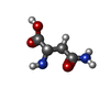  Type: L-peptide linking / Mass: 132.118 Da / Num. of mol.: 4 / Source method: obtained synthetically / Formula: C4H8N2O3 Type: L-peptide linking / Mass: 132.118 Da / Num. of mol.: 4 / Source method: obtained synthetically / Formula: C4H8N2O3#3: Chemical | ChemComp-EDO /   Mass: 62.068 Da / Num. of mol.: 9 / Source method: obtained synthetically / Formula: C2H6O2 Mass: 62.068 Da / Num. of mol.: 9 / Source method: obtained synthetically / Formula: C2H6O2#4: Water | ChemComp-HOH / |  Mass: 18.015 Da / Num. of mol.: 320 / Source method: isolated from a natural source / Formula: H2O Mass: 18.015 Da / Num. of mol.: 320 / Source method: isolated from a natural source / Formula: H2O |
|---|
-Experimental details
-Experiment
| Experiment | Method: X-RAY DIFFRACTION |
|---|
- Sample preparation
| Crystal | Density Matthews: 1.49 Å3/Da / Density % sol: 17.31 % |
|---|---|
| Crystal grow | Temperature: 293 K / Method: vapor diffusion, hanging drop / pH: 7 / Details: 0.1 M HEPES pH 7.0, 12-15% PEG 4000 |
-Data collection
| Diffraction | Mean temperature: 100 K |
|---|---|
| Diffraction source | Source: SYNCHROTRON / Site: APS  / Beamline: 21-ID-G / Wavelength: 0.97857 Å / Beamline: 21-ID-G / Wavelength: 0.97857 Å |
| Detector | Type: MARMOSAIC 300 mm CCD / Detector: CCD / Date: Apr 23, 2015 |
| Radiation | Monochromator: DIAMOND / Protocol: SINGLE WAVELENGTH / Monochromatic (M) / Laue (L): M / Scattering type: x-ray |
| Radiation wavelength | Wavelength: 0.97857 Å / Relative weight: 1 |
| Reflection | Resolution: 2.39→30 Å / Num. obs: 57320 / % possible obs: 95.5 % / Redundancy: 3.2 % / Rsym value: 0.12 / Net I/σ(I): 9.1 |
| Reflection shell | Resolution: 2.39→2.53 Å / Redundancy: 3.1 % / % possible all: 93.8 |
- Processing
| Software |
| ||||||||||||||||||||||||||||||||||||||||||||||||||||||||||||||||||||||||||||||||||||||||||||||||||||||||||||||||||||||||||||||||||||||||||||||||||||||||||||||||||||||||||||||||||||||
|---|---|---|---|---|---|---|---|---|---|---|---|---|---|---|---|---|---|---|---|---|---|---|---|---|---|---|---|---|---|---|---|---|---|---|---|---|---|---|---|---|---|---|---|---|---|---|---|---|---|---|---|---|---|---|---|---|---|---|---|---|---|---|---|---|---|---|---|---|---|---|---|---|---|---|---|---|---|---|---|---|---|---|---|---|---|---|---|---|---|---|---|---|---|---|---|---|---|---|---|---|---|---|---|---|---|---|---|---|---|---|---|---|---|---|---|---|---|---|---|---|---|---|---|---|---|---|---|---|---|---|---|---|---|---|---|---|---|---|---|---|---|---|---|---|---|---|---|---|---|---|---|---|---|---|---|---|---|---|---|---|---|---|---|---|---|---|---|---|---|---|---|---|---|---|---|---|---|---|---|---|---|---|---|
| Refinement | Method to determine structure: MOLECULAR REPLACEMENT Starting model: 4r8l Resolution: 2.39→30 Å / Cor.coef. Fo:Fc: 0.953 / Cor.coef. Fo:Fc free: 0.927 / SU B: 11.839 / SU ML: 0.244 / Cross valid method: THROUGHOUT / ESU R: 0.537 / ESU R Free: 0.268 / Stereochemistry target values: MAXIMUM LIKELIHOOD / Details: HYDROGENS HAVE BEEN ADDED IN THE RIDING POSITIONS
| ||||||||||||||||||||||||||||||||||||||||||||||||||||||||||||||||||||||||||||||||||||||||||||||||||||||||||||||||||||||||||||||||||||||||||||||||||||||||||||||||||||||||||||||||||||||
| Solvent computation | Ion probe radii: 0.8 Å / Shrinkage radii: 0.8 Å / VDW probe radii: 1.2 Å / Solvent model: MASK | ||||||||||||||||||||||||||||||||||||||||||||||||||||||||||||||||||||||||||||||||||||||||||||||||||||||||||||||||||||||||||||||||||||||||||||||||||||||||||||||||||||||||||||||||||||||
| Displacement parameters | Biso mean: 40.279 Å2
| ||||||||||||||||||||||||||||||||||||||||||||||||||||||||||||||||||||||||||||||||||||||||||||||||||||||||||||||||||||||||||||||||||||||||||||||||||||||||||||||||||||||||||||||||||||||
| Refinement step | Cycle: 1 / Resolution: 2.39→30 Å
| ||||||||||||||||||||||||||||||||||||||||||||||||||||||||||||||||||||||||||||||||||||||||||||||||||||||||||||||||||||||||||||||||||||||||||||||||||||||||||||||||||||||||||||||||||||||
| Refine LS restraints |
|