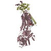
 Movie
Movie Controller

[English] 日本語
 Yorodumi
- PDB-5avw: Kinetics by X-ray crystallography: Tl+-substitution of bound K+ i... -
+ Open data

- Basic information
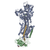
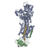
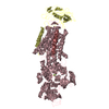
| Entry | Database: PDB / ID: 5avw | |||||||||||||||
|---|---|---|---|---|---|---|---|---|---|---|---|---|---|---|---|---|
| Title | Kinetics by X-ray crystallography: Tl+-substitution of bound K+ in the E2.MgF42-.2K+ crystal after 16.5 min | |||||||||||||||
 Components Components |
| |||||||||||||||
 Keywords Keywords | HYDROLASE/TRANSPORT PROTEIN / MEMBRANE PROTEIN / ION PUMP / ATPASE / K+ BINDING / HALOACID DEHYDROGENEASE SUPERFAMILY / PHOSPHATE ANALOGUE / ATP-BINDING / HYDROLASE / ION TRANSPORT / NUCLEOTIDE-BINDING / PHOSPHOPROTEIN / HYDROLASE-TRANSPORT PROTEIN COMPLEX / KINETICS | |||||||||||||||
| Function / homology |  Function and homology information Function and homology informationregulation of monoatomic ion transport / P-type sodium:potassium-exchanging transporter activity / sodium:potassium-exchanging ATPase complex / sodium ion export across plasma membrane / intracellular sodium ion homeostasis / potassium ion import across plasma membrane / intracellular potassium ion homeostasis / ATPase activator activity / sodium channel regulator activity / monoatomic ion transport ...regulation of monoatomic ion transport / P-type sodium:potassium-exchanging transporter activity / sodium:potassium-exchanging ATPase complex / sodium ion export across plasma membrane / intracellular sodium ion homeostasis / potassium ion import across plasma membrane / intracellular potassium ion homeostasis / ATPase activator activity / sodium channel regulator activity / monoatomic ion transport / proton transmembrane transport / ATP hydrolysis activity / ATP binding / metal ion binding / membrane / plasma membrane Similarity search - Function | |||||||||||||||
| Biological species |  Squalus acanthias (spiny dogfish) Squalus acanthias (spiny dogfish) | |||||||||||||||
| Method |  X-RAY DIFFRACTION / SYNCHROTRON / MOLECULAR REPLACEMENT / Resolution: 2.6 Å X-RAY DIFFRACTION / SYNCHROTRON / MOLECULAR REPLACEMENT / Resolution: 2.6 Å | |||||||||||||||
 Authors Authors | Ogawa, H. / Cornelius, F. / Hirata, A. / Toyoshima, C. | |||||||||||||||
| Funding support |  Japan, Japan,  Denmark, 4items Denmark, 4items
| |||||||||||||||
 Citation Citation | Journal: Nat Commun / Year: 2015 Title: Sequential substitution of K(+) bound to Na(+),K(+)-ATPase visualized by X-ray crystallography. Authors: Ogawa, H. / Cornelius, F. / Hirata, A. / Toyoshima, C. | |||||||||||||||
| History |
|
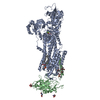
- Structure visualization
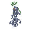
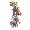
| Structure viewer | Molecule: MolmilJmol/JSmol |
|---|
- Downloads & links
-Download
| PDBx/mmCIF format | 5avw.cif.gz | 270.5 KB | Display | PDBx/mmCIF format |
|---|---|---|---|---|
| PDB format | pdb5avw.ent.gz | 209.2 KB | Display | PDB format |
| PDBx/mmJSON format | 5avw.json.gz | Tree view | PDBx/mmJSON format | |
| Others |  Other downloads Other downloads |
-Validation report
| Arichive directory | https://data.pdbj.org/pub/pdb/validation_reports/av/5avwftp://data.pdbj.org/pub/pdb/validation_reports/av/5avw | HTTPS FTP |
|---|
-Related structure data
| Related structure data |  5avqC  5avrC  5avsC  5avtC  5avuC  5avvC  5avxC  5avyC  5avzC  5aw0C  5aw1C  5aw2C  5aw3C  5aw4C  5aw5C  5aw6C  5aw7C  5aw8C  5aw9C  2zxeS C: citing same article ( S: Starting model for refinement |
|---|---|
| Similar structure data |


-Links
 PDBj
PDBj


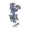
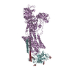
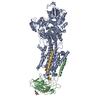
- Assembly
| Deposited unit | 
| ||||||||
|---|---|---|---|---|---|---|---|---|---|
| 1 |
| ||||||||
| Unit cell |
|
-Components
-Protein , 3 types, 3 molecules ABG
| #1: Protein | Mass: 113309.891 Da / Num. of mol.: 1 / Source method: isolated from a natural source / Source: (natural) Squalus acanthias (spiny dogfish) / References: UniProt: Q4H132 |
|---|---|
| #2: Protein | Mass: 35176.125 Da / Num. of mol.: 1 / Source method: isolated from a natural source / Source: (natural) Squalus acanthias (spiny dogfish) / References: UniProt: C4IX13 |
| #3: Protein | Mass: 8225.446 Da / Num. of mol.: 1 / Source method: isolated from a natural source / Source: (natural) Squalus acanthias (spiny dogfish) / References: UniProt: Q70Q12 |
-Sugars , 2 types, 2 molecules 
| #4: Polysaccharide | 2-acetamido-2-deoxy-beta-D-glucopyranose-(1-4)-2-acetamido-2-deoxy-beta-D-glucopyranose Source method: isolated from a genetically manipulated source |
|---|---|
| #10: Sugar | ChemComp-NAG /  Type: D-saccharide, beta linking / Mass: 221.208 Da / Num. of mol.: 1 Type: D-saccharide, beta linking / Mass: 221.208 Da / Num. of mol.: 1Source method: isolated from a genetically manipulated source Formula: C8H15NO6 |
-Non-polymers , 6 types, 8 molecules 


| #5: Chemical | ChemComp-MF4 /  Mass: 100.299 Da / Num. of mol.: 1 / Source method: obtained synthetically / Formula: F4Mg Mass: 100.299 Da / Num. of mol.: 1 / Source method: obtained synthetically / Formula: F4Mg | ||||||
|---|---|---|---|---|---|---|---|
| #6: Chemical | ChemComp-MG /  Mass: 24.305 Da / Num. of mol.: 1 / Source method: obtained synthetically / Formula: Mg Mass: 24.305 Da / Num. of mol.: 1 / Source method: obtained synthetically / Formula: Mg | ||||||
| #7: Chemical |  Mass: 204.383 Da / Num. of mol.: 3 / Source method: obtained synthetically / Formula: Tl Mass: 204.383 Da / Num. of mol.: 3 / Source method: obtained synthetically / Formula: Tl#8: Chemical | ChemComp-K / |  Mass: 39.098 Da / Num. of mol.: 1 / Source method: obtained synthetically / Formula: K Mass: 39.098 Da / Num. of mol.: 1 / Source method: obtained synthetically / Formula: K#9: Chemical | ChemComp-CLR / |  Mass: 386.654 Da / Num. of mol.: 1 / Source method: obtained synthetically / Formula: C27H46O Mass: 386.654 Da / Num. of mol.: 1 / Source method: obtained synthetically / Formula: C27H46O#11: Water | ChemComp-HOH / | Mass: 18.015 Da / Num. of mol.: 1 / Source method: isolated from a natural source / Formula: H2O |
-Details
| Has protein modification | Y |
|---|
-Experimental details
-Experiment
| Experiment | Method: X-RAY DIFFRACTION |
|---|
- Sample preparation
| Crystal | Density Matthews: 2.86 Å3/Da / Density % sol: 57.02 % |
|---|---|
| Crystal grow | Temperature: 298 K / Method: microdialysis / pH: 7 Details: PEG 3000, MPD, potassium acetate, potassium chloride, magnesium chloride, potassium fluoride, MES/TRIS |
-Data collection
| Diffraction | Mean temperature: 100 K |
|---|---|
| Diffraction source | Source: SYNCHROTRON / Site: SPring-8 / Beamline: BL41XU / Wavelength: 0.9785 Å |
| Detector | Type: RAYONIX MX225HE / Detector: CCD / Date: Oct 10, 2009 |
| Radiation | Protocol: SINGLE WAVELENGTH / Monochromatic (M) / Laue (L): M / Scattering type: x-ray |
| Radiation wavelength | Wavelength: 0.9785 Å / Relative weight: 1 |
| Reflection | Resolution: 2.6→50 Å / Num. obs: 54317 / % possible obs: 97.9 % / Redundancy: 4.1 % / Biso Wilson estimate: 29.8 Å2 / Net I/σ(I): 22.9 |
| Reflection shell | Resolution: 2.6→2.67 Å |
- Processing
| Software |
| ||||||||||||||||||||||||||||||||||||||||||||||||||||||||||||||||||||||||||||||||
|---|---|---|---|---|---|---|---|---|---|---|---|---|---|---|---|---|---|---|---|---|---|---|---|---|---|---|---|---|---|---|---|---|---|---|---|---|---|---|---|---|---|---|---|---|---|---|---|---|---|---|---|---|---|---|---|---|---|---|---|---|---|---|---|---|---|---|---|---|---|---|---|---|---|---|---|---|---|---|---|---|---|
| Refinement | Method to determine structure: MOLECULAR REPLACEMENT Starting model: 2ZXE Resolution: 2.6→14.98 Å / Rfactor Rfree error: 0.007 / Data cutoff high absF: 2944211.32 / Data cutoff low absF: 0 / Isotropic thermal model: RESTRAINED / Cross valid method: THROUGHOUT / σ(F): 3.5 / Details: BULK SOLVENT MODEL USED, Rigid body refinement
| ||||||||||||||||||||||||||||||||||||||||||||||||||||||||||||||||||||||||||||||||
| Solvent computation | Solvent model: FLAT MODEL / Bsol: 49.93 Å2 / ksol: 0.35 e/Å3 | ||||||||||||||||||||||||||||||||||||||||||||||||||||||||||||||||||||||||||||||||
| Displacement parameters | Biso mean: 83.5 Å2
| ||||||||||||||||||||||||||||||||||||||||||||||||||||||||||||||||||||||||||||||||
| Refine analyze |
| ||||||||||||||||||||||||||||||||||||||||||||||||||||||||||||||||||||||||||||||||
| Refinement step | Cycle: LAST / Resolution: 2.6→14.98 Å
| ||||||||||||||||||||||||||||||||||||||||||||||||||||||||||||||||||||||||||||||||
| Refine LS restraints |
| ||||||||||||||||||||||||||||||||||||||||||||||||||||||||||||||||||||||||||||||||
| LS refinement shell | Resolution: 2.6→2.76 Å / Rfactor Rfree error: 0.036 / Total num. of bins used: 6
| ||||||||||||||||||||||||||||||||||||||||||||||||||||||||||||||||||||||||||||||||
| Xplor file |
|
