[English] 日本語
Yorodumi
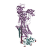
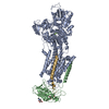
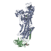
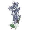
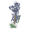
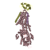
- PDB-3a3y: Crystal structure of the sodium-potassium pump with bound potassi... -

+
Open data


ID or keywords:

Loading...

-
Basic information

Entry
Database: PDB / ID: 3a3y
TitleCrystal structure of the sodium-potassium pump with bound potassium and ouabain
Components
  • NA+,K+-ATPASE BETA SUBUNIT
  • Na, K-ATPase alpha subunit
  • Phospholemman-like protein
KeywordsHYDROLASE/TRANSPORT PROTEIN / MEMBRANE PROTEIN / ION PUMP / ATPASE / K+ BINDING / OUABAIN BINDING / HALOACID DEHYDROGENEASE SUPERFAMILY / PHOSPHATE ANALOGUE / ATP-BINDING / HYDROLASE / ION TRANSPORT / NUCLEOTIDE-BINDING / PHOSPHOPROTEIN / HYDROLASE-TRANSPORT PROTEIN COMPLEX / Membrane / Transmembrane / Transport
Function / homology
Function and homology information


regulation of monoatomic ion transport / P-type potassium transmembrane transporter activity / sodium:potassium-exchanging ATPase complex / ion channel regulator activity / sodium ion transport / monoatomic ion transport / potassium ion transport / ATP hydrolysis activity / ATP binding / membrane ...regulation of monoatomic ion transport / P-type potassium transmembrane transporter activity / sodium:potassium-exchanging ATPase complex / ion channel regulator activity / sodium ion transport / monoatomic ion transport / potassium ion transport / ATP hydrolysis activity / ATP binding / membrane / metal ion binding / plasma membrane
Similarity search - Function
Na, k-atpase alpha subunit. / : / Ion-transport regulator, FXYD motif / ATP1G1/PLM/MAT8 family / FXYD family signature. / Calcium-transporting ATPase, transmembrane domain / Calcium-transporting ATPase, transmembrane domain / Calcium-transporting ATPase, cytoplasmic transduction domain A / Calcium-transporting ATPase, cytoplasmic transduction domain A / Sodium/potassium-transporting ATPase subunit beta ...Na, k-atpase alpha subunit. / : / Ion-transport regulator, FXYD motif / ATP1G1/PLM/MAT8 family / FXYD family signature. / Calcium-transporting ATPase, transmembrane domain / Calcium-transporting ATPase, transmembrane domain / Calcium-transporting ATPase, cytoplasmic transduction domain A / Calcium-transporting ATPase, cytoplasmic transduction domain A / Sodium/potassium-transporting ATPase subunit beta / Sodium/potassium-transporting ATPase subunit beta superfamily / Sodium / potassium ATPase beta chain / Sodium and potassium ATPases beta subunits signature 1. / Sodium and potassium ATPases beta subunits signature 2. / Calcium-transporting ATPase, cytoplasmic domain N / Calcium-transporting ATPase, cytoplasmic domain N / P-type ATPase subfamily IIC, subunit alpha / Single alpha-helices involved in coiled-coils or other helix-helix interfaces - #170 / Cation-transporting P-type ATPase, C-terminal / Cation transporting ATPase, C-terminus / Cation transporter/ATPase, N-terminus / Cation-transporting P-type ATPase, N-terminal / Cation transporter/ATPase, N-terminus / Cation transport ATPase (P-type) / E1-E2 ATPase / P-type ATPase, haloacid dehalogenase domain / P-type ATPase, phosphorylation site / P-type ATPase, cytoplasmic domain N / E1-E2 ATPases phosphorylation site. / P-type ATPase, A domain superfamily / P-type ATPase / P-type ATPase, transmembrane domain superfamily / haloacid dehalogenase-like hydrolase / HAD superfamily/HAD-like / HAD superfamily / HAD-like superfamily / Single alpha-helices involved in coiled-coils or other helix-helix interfaces / Distorted Sandwich / Up-down Bundle / Immunoglobulin-like / Sandwich / Rossmann fold / 3-Layer(aba) Sandwich / Mainly Beta / Mainly Alpha / Alpha Beta
Similarity search - Domain/homology
CHOLESTEROL / : / TETRAFLUOROMAGNESATE(2-) / OUABAIN / Sodium/potassium-transporting ATPase subunit beta / Sodium/potassium-transporting ATPase subunit alpha / FXYD domain-containing ion transport regulator
Similarity search - Component
Biological speciesSqualus acanthias (spiny dogfish)
SQUALUS ACANTHIAS (spiny dogfish)
MethodX-RAY DIFFRACTION / SYNCHROTRON / MOLECULAR REPLACEMENT / Resolution: 2.8 Å
AuthorsOgawa, H. / Shinoda, T. / Cornelius, F. / Toyoshima, C.
CitationJournal: Proc.Natl.Acad.Sci.USA / Year: 2009
Title: Crystal structure of the sodium-potassium pump (Na+,K+-ATPase) with bound potassium and ouabain.
Authors: Ogawa, H. / Shinoda, T. / Cornelius, F. / Toyoshima, C.
History
DepositionJun 23, 2009Deposition site: PDBJ / Processing site: PDBJ
Revision 1.0Sep 8, 2009Provider: repository / Type: Initial release
Revision 1.1Jul 13, 2011Group: Advisory / Version format compliance
Revision 1.2Jul 29, 2020Group: Data collection / Derived calculations / Structure summary
Category: chem_comp / entity ...chem_comp / entity / pdbx_chem_comp_identifier / pdbx_entity_nonpoly / struct_site / struct_site_gen
Item: _chem_comp.name / _chem_comp.type ..._chem_comp.name / _chem_comp.type / _entity.pdbx_description / _pdbx_entity_nonpoly.name
Description: Carbohydrate remediation / Provider: repository / Type: Remediation

-
Structure visualization

Structure viewerMolecule:
MolmilJmol/JSmol

Downloads & links

-
Assembly

Deposited unit
A: Na, K-ATPase alpha subunit
B: NA+,K+-ATPASE BETA SUBUNIT
G: Phospholemman-like protein
hetero molecules


Theoretical massNumber of molelcules
Total (without water)158,58713
Polymers156,7103
Non-polymers1,87710
Water1,18966
1


  • Idetical with deposited unit
  • defined by author&software
TypeNameSymmetry operationNumber
identity operation1_555x,y,z1
Buried area6490 Å2
ΔGint-37 kcal/mol
Surface area56180 Å2
MethodPISA
Unit cell
Length a, b, c (Å)222.884, 50.720, 163.342
Angle α, β, γ (deg.)90.00, 104.63, 90.00
Int Tables number5
Space group name H-MC121

-
Components

-
Protein , 3 types, 3 molecules ABG

#1: Protein Na, K-ATPase alpha subunit


Mass: 113309.891 Da / Num. of mol.: 1 / Source method: isolated from a natural source / Source: (natural) Squalus acanthias (spiny dogfish) / References: UniProt: Q4H132
#2: Protein NA+,K+-ATPASE BETA SUBUNIT


Mass: 35175.141 Da / Num. of mol.: 1 / Source method: isolated from a natural source / Source: (natural) SQUALUS ACANTHIAS (spiny dogfish) / References: UniProt: C4IX13*PLUS
#3: Protein Phospholemman-like protein


Mass: 8225.446 Da / Num. of mol.: 1 / Source method: isolated from a natural source / Source: (natural) Squalus acanthias (spiny dogfish) / References: UniProt: Q70Q12

-
Sugars , 1 types, 3 molecules

#9: Sugar ChemComp-NAG / 2-acetamido-2-deoxy-beta-D-glucopyranose / N-Acetylglucosamine


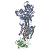
Type: D-saccharide, beta linking / Mass: 221.208 Da / Num. of mol.: 3
Source method: isolated from a genetically manipulated source
Formula: C8H15NO6
IdentifierTypeProgram
DGlcpNAcbCONDENSED IUPAC CARBOHYDRATE SYMBOLGMML 1.0
N-acetyl-b-D-glucopyranosamineCOMMON NAMEGMML 1.0
b-D-GlcpNAcIUPAC CARBOHYDRATE SYMBOLPDB-CARE 1.0
GlcNAcSNFG CARBOHYDRATE SYMBOLGMML 1.0

-
Non-polymers , 6 types, 73 molecules

#4: Chemical ChemComp-MF4 / TETRAFLUOROMAGNESATE(2-) / MAGNESIUMTETRAFLUORIDE


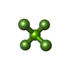
Mass: 100.299 Da / Num. of mol.: 1 / Source method: obtained synthetically / Formula: F4Mg
#5: Chemical ChemComp-MG / MAGNESIUM ION


Mass: 24.305 Da / Num. of mol.: 1 / Source method: obtained synthetically / Formula: Mg
#6: Chemical ChemComp-K / POTASSIUM ION


Mass: 39.098 Da / Num. of mol.: 3 / Source method: obtained synthetically / Formula: K
#7: Chemical ChemComp-OBN / OUABAIN / Ouabain


Mass: 584.652 Da / Num. of mol.: 1 / Source method: obtained synthetically / Formula: C29H44O12
#8: Chemical ChemComp-CLR / CHOLESTEROL / Cholesterol


Mass: 386.654 Da / Num. of mol.: 1 / Source method: obtained synthetically / Formula: C27H46O
#10: Water ChemComp-HOH / water / Water


Mass: 18.015 Da / Num. of mol.: 66 / Source method: isolated from a natural source / Formula: H2O

-
Details

Sequence detailsA SEQUENCE DATABASE REFERENCE FOR THE CHAIN B DOES NOT CURRENTLY EXIST IN UNIPROT.

-
Experimental details

-
Experiment

ExperimentMethod: X-RAY DIFFRACTION / Number of used crystals: 2

-
Sample preparation

CrystalDensity Matthews: 2.85 Å3/Da / Density % sol: 56.85 %

-
Data collection

DiffractionMean temperature: 100 K
Diffraction sourceSource: SYNCHROTRON / Site: SPring-8 / Beamline: BL41XU / Wavelength: 0.9 Å
DetectorType: RAYONIX MX225HE / Detector: CCD
RadiationProtocol: SINGLE WAVELENGTH / Monochromatic (M) / Laue (L): M / Scattering type: x-ray
Radiation wavelengthWavelength: 0.9 Å / Relative weight: 1
ReflectionResolution: 2.8→50 Å / Num. obs: 44358 / % possible obs: 99.9 % / Redundancy: 10.4 % / Rmerge(I) obs: 0.045 / Net I/σ(I): 30.3
Reflection shellResolution: 2.8→2.9 Å / Redundancy: 8.2 % / Rmerge(I) obs: 0.388 / Mean I/σ(I) obs: 3.2 / Num. unique all: 4350 / % possible all: 100

-
Processing

Software
NameVersionClassification
REFMAC5.5.0072refinement
DENZOdata reduction
SCALEPACKdata scaling
CNSphasing
RefinementMethod to determine structure: MOLECULAR REPLACEMENT / Resolution: 2.8→15 Å / Cor.coef. Fo:Fc: 0.921 / Cor.coef. Fo:Fc free: 0.878 / SU B: 40.906 / SU ML: 0.361 / TLS residual ADP flag: LIKELY RESIDUAL / Cross valid method: THROUGHOUT / ESU R: 1.859 / ESU R Free: 0.407 / Stereochemistry target values: MAXIMUM LIKELIHOOD / Details: HYDROGENS HAVE BEEN ADDED IN THE RIDING POSITIONS
RfactorNum. reflection% reflectionSelection details
Rfree0.2928 1333 3 %RANDOM
Rwork0.24715 ---
obs0.24855 42690 99.95 %-
Solvent computationIon probe radii: 0.8 Å / Shrinkage radii: 0.8 Å / VDW probe radii: 1.4 Å / Solvent model: MASK
Displacement parametersBiso mean: 63.075 Å2
Baniso -1Baniso -2Baniso -3
1-4.42 Å20 Å2-1.02 Å2
2---1.29 Å20 Å2
3----3.65 Å2
Refinement stepCycle: LAST / Resolution: 2.8→15 Å
ProteinNucleic acidLigandSolventTotal
Num. atoms10122 0 120 66 10308
Refine LS restraints
Refine-IDTypeDev idealDev ideal targetNumber
X-RAY DIFFRACTIONr_bond_refined_d0.0070.02210456
X-RAY DIFFRACTIONr_bond_other_d
X-RAY DIFFRACTIONr_angle_refined_deg1.211.98214190
X-RAY DIFFRACTIONr_angle_other_deg
X-RAY DIFFRACTIONr_dihedral_angle_1_deg5.67251290
X-RAY DIFFRACTIONr_dihedral_angle_2_deg36.07124.089450
X-RAY DIFFRACTIONr_dihedral_angle_3_deg17.845151783
X-RAY DIFFRACTIONr_dihedral_angle_4_deg16.471563
X-RAY DIFFRACTIONr_chiral_restr0.0790.21620
X-RAY DIFFRACTIONr_gen_planes_refined0.0040.0217787
X-RAY DIFFRACTIONr_gen_planes_other
X-RAY DIFFRACTIONr_nbd_refined
X-RAY DIFFRACTIONr_nbd_other
X-RAY DIFFRACTIONr_nbtor_refined
X-RAY DIFFRACTIONr_nbtor_other
X-RAY DIFFRACTIONr_xyhbond_nbd_refined
X-RAY DIFFRACTIONr_xyhbond_nbd_other
X-RAY DIFFRACTIONr_metal_ion_refined
X-RAY DIFFRACTIONr_metal_ion_other
X-RAY DIFFRACTIONr_symmetry_vdw_refined
X-RAY DIFFRACTIONr_symmetry_vdw_other
X-RAY DIFFRACTIONr_symmetry_hbond_refined
X-RAY DIFFRACTIONr_symmetry_hbond_other
X-RAY DIFFRACTIONr_symmetry_metal_ion_refined
X-RAY DIFFRACTIONr_symmetry_metal_ion_other
X-RAY DIFFRACTIONr_mcbond_it0.9281.56435
X-RAY DIFFRACTIONr_mcbond_other
X-RAY DIFFRACTIONr_mcangle_it1.519210404
X-RAY DIFFRACTIONr_scbond_it1.36834021
X-RAY DIFFRACTIONr_scangle_it2.0724.53786
X-RAY DIFFRACTIONr_rigid_bond_restr
X-RAY DIFFRACTIONr_sphericity_free
X-RAY DIFFRACTIONr_sphericity_bonded
LS refinement shellResolution: 2.8→2.87 Å / Total num. of bins used: 20
RfactorNum. reflection% reflection
Rfree0.421 81 -
Rwork0.338 3052 -
obs--99.9 %
Refinement TLS params.

Method: refined / Refine-ID: X-RAY DIFFRACTION

IDL112)L122)L132)L222)L232)L332)S11 (Å °)S12 (Å °)S13 (Å °)S21 (Å °)S22 (Å °)S23 (Å °)S31 (Å °)S32 (Å °)S33 (Å °)T112)T122)T132)T222)T232)T332)Origin x (Å)Origin y (Å)Origin z (Å)
11.1590.8304-0.03031.86670.42571.09910.0293-0.0161-0.01580.16190.0245-0.08740.15640.1763-0.05380.03450.0287-0.04080.16550.0290.1838150.520640.160465.9105
20.9625-0.3765-0.26152.52430.583.6905-0.2075-0.0482-0.09130.3378-0.06170.4081-0.0674-0.30960.26920.08480.01610.0760.0855-0.02020.2309112.189639.003177.473
30.32260.1193-0.03920.9841-0.43751.2709-0.01030.02860.0299-0.0431-0.00070.04550.14350.0650.0110.0420.0188-0.02930.1280.00240.1878132.16218.977954.8715
40.0635-0.081-0.02940.27720.20121.24160.00770.02240.03940.0202-0.0301-0.05370.08340.11870.02240.09470.0182-0.00490.18280.02550.1355129.719314.69796.2859
50.3982-0.0639-0.97752.24133.56347.6844-0.0263-0.0560.3384-0.0230.25720.3431-0.10570.7801-0.23090.2624-0.1455-0.0570.61580.05720.3716149.105828.506714.75
61.9558-0.2429-0.24661.0399-0.25392.8020.18150.50680.2291-0.4325-0.2479-0.1363-0.06010.31960.06640.34740.09780.11770.45830.12390.0663142.427920.6871-37.3312
Refinement TLS group
IDRefine-IDRefine TLS-IDAuth asym-IDAuth seq-ID
1X-RAY DIFFRACTION1A32 - 81
2X-RAY DIFFRACTION1A157 - 276
3X-RAY DIFFRACTION2A384 - 594
4X-RAY DIFFRACTION3A355 - 383
5X-RAY DIFFRACTION3A595 - 754
6X-RAY DIFFRACTION4A277 - 354
7X-RAY DIFFRACTION4A755 - 1023
8X-RAY DIFFRACTION4B35 - 72
9X-RAY DIFFRACTION4G8 - 46
10X-RAY DIFFRACTION5A82 - 156
11X-RAY DIFFRACTION6B73 - 305

+
About Yorodumi

-
News

-
Feb 9, 2022. New format data for meta-information of EMDB entries

New format data for meta-information of EMDB entries

  • Version 3 of the EMDB header file is now the official format.
  • The previous official version 1.9 will be removed from the archive.

Related info.:EMDB header

External links:wwPDB to switch to version 3 of the EMDB data model

-
Aug 12, 2020. Covid-19 info

Covid-19 info

URL: https://pdbj.org/emnavi/covid19.php

New page: Covid-19 featured information page in EM Navigator.

Related info.:Covid-19 info / Mar 5, 2020. Novel coronavirus structure data

+
Mar 5, 2020. Novel coronavirus structure data

Novel coronavirus structure data

Related info.:Yorodumi Speices / Aug 12, 2020. Covid-19 info

External links:COVID-19 featured content - PDBj / Molecule of the Month (242):Coronavirus Proteases

+
Jan 31, 2019. EMDB accession codes are about to change! (news from PDBe EMDB page)

EMDB accession codes are about to change! (news from PDBe EMDB page)

  • The allocation of 4 digits for EMDB accession codes will soon come to an end. Whilst these codes will remain in use, new EMDB accession codes will include an additional digit and will expand incrementally as the available range of codes is exhausted. The current 4-digit format prefixed with “EMD-” (i.e. EMD-XXXX) will advance to a 5-digit format (i.e. EMD-XXXXX), and so on. It is currently estimated that the 4-digit codes will be depleted around Spring 2019, at which point the 5-digit format will come into force.
  • The EM Navigator/Yorodumi systems omit the EMD- prefix.

Related info.:Q: What is EMD? / ID/Accession-code notation in Yorodumi/EM Navigator

External links:EMDB Accession Codes are Changing Soon! / Contact to PDBj

+
Jul 12, 2017. Major update of PDB

Major update of PDB

  • wwPDB released updated PDB data conforming to the new PDBx/mmCIF dictionary.
  • This is a major update changing the version number from 4 to 5, and with Remediation, in which all the entries are updated.
  • In this update, many items about electron microscopy experimental information are reorganized (e.g. em_software).
  • Now, EM Navigator and Yorodumi are based on the updated data.

External links:wwPDB Remediation / Enriched Model Files Conforming to OneDep Data Standards Now Available in the PDB FTP Archive

-
Yorodumi

Thousand views of thousand structures

  • Yorodumi is a browser for structure data from EMDB, PDB, SASBDB, etc.
  • This page is also the successor to EM Navigator detail page, and also detail information page/front-end page for Omokage search.
  • The word "yorodu" (or yorozu) is an old Japanese word meaning "ten thousand". "mi" (miru) is to see.

Related info.:EMDB / PDB / SASBDB / Comparison of 3 databanks / Yorodumi Search / Aug 31, 2016. New EM Navigator & Yorodumi / Yorodumi Papers / Jmol/JSmol / Function and homology information / Changes in new EM Navigator and Yorodumi

Read more