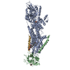
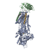
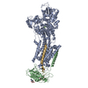
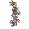
: / Ion homeostasis / Ion transport by P-type ATPases / Na+/K+-exchanging ATPase / regulation of monoatomic ion transport / positive regulation of sodium ion export across plasma membrane / positive regulation of potassium ion import across plasma membrane / P-type sodium:potassium-exchanging transporter activity / sodium ion binding / sodium:potassium-exchanging ATPase complex ...: / Ion homeostasis / Ion transport by P-type ATPases / Na+/K+-exchanging ATPase / regulation of monoatomic ion transport / positive regulation of sodium ion export across plasma membrane / positive regulation of potassium ion import across plasma membrane / P-type sodium:potassium-exchanging transporter activity / sodium ion binding / sodium:potassium-exchanging ATPase complex / membrane repolarization / establishment or maintenance of transmembrane electrochemical gradient / sodium ion export across plasma membrane / regulation of calcium ion transmembrane transport / ion channel regulator activity / intracellular sodium ion homeostasis / positive regulation of potassium ion transmembrane transport / relaxation of cardiac muscle / regulation of cardiac muscle contraction by calcium ion signaling / positive regulation of sodium ion transmembrane transport / organelle membrane / potassium ion binding / potassium ion import across plasma membrane / ATPase activator activity / intracellular potassium ion homeostasis / intercalated disc / lateral plasma membrane / transporter activator activity / sperm flagellum / ATP metabolic process / regulation of sodium ion transport / cardiac muscle contraction / T-tubule / proton transmembrane transport / protein localization to plasma membrane / sarcolemma / transmembrane transport / intracellular calcium ion homeostasis / melanosome / ATPase binding / regulation of gene expression / basolateral plasma membrane / protein-macromolecule adaptor activity / cell adhesion / apical plasma membrane / protein stabilization / innate immune response / axon / protein kinase binding / ATP hydrolysis activity / ATP binding / membrane / plasma membrane Similarity search - Function
In the structure databanks used in Yorodumi, some data are registered as the other names, "COVID-19 virus" and "2019-nCoV". Here are the details of the virus and the list of structure data.
Jan 31, 2019. EMDB accession codes are about to change! (news from PDBe EMDB page)
EMDB accession codes are about to change! (news from PDBe EMDB page)
The allocation of 4 digits for EMDB accession codes will soon come to an end. Whilst these codes will remain in use, new EMDB accession codes will include an additional digit and will expand incrementally as the available range of codes is exhausted. The current 4-digit format prefixed with “EMD-” (i.e. EMD-XXXX) will advance to a 5-digit format (i.e. EMD-XXXXX), and so on. It is currently estimated that the 4-digit codes will be depleted around Spring 2019, at which point the 5-digit format will come into force.
The EM Navigator/Yorodumi systems omit the EMD- prefix.
Related info.:Q: What is EMD? / ID/Accession-code notation in Yorodumi/EM Navigator
Yorodumi is a browser for structure data from EMDB, PDB, SASBDB, etc.
This page is also the successor to EM Navigator detail page, and also detail information page/front-end page for Omokage search.
The word "yorodu" (or yorozu) is an old Japanese word meaning "ten thousand". "mi" (miru) is to see.
Related info.:EMDB / PDB / SASBDB / Comparison of 3 databanks / Yorodumi Search / Aug 31, 2016. New EM Navigator & Yorodumi / Yorodumi Papers / Jmol/JSmol / Function and homology information / Changes in new EM Navigator and Yorodumi
 Movie
Movie Controller

 Yorodumi
 Open data
 Basic information
 Components
 Keywords
 Function and homology information
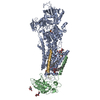
 X-RAY DIFFRACTION /
  Authors
 Citation
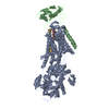
 Structure visualization
 Downloads & links
 Other downloads
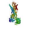


 PDBj
PDBj

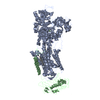
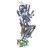
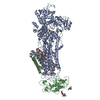
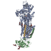
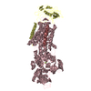
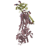
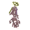
 Assembly
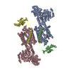

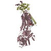
 Mass: 584.652 Da / Num. of mol.: 2 / Source method: obtained synthetically / Formula: C29H44O12
 Mass: 24.305 Da / Num. of mol.: 2 / Source method: obtained synthetically / Formula: Mg
 Sample preparation
 / Beamline: ID29 / Wavelength: 0.976 Å
 Processing