 Movie
Movie Controller


+ Open data

- Basic information
| Entry | Database: PDB / ID: 5ae0 | |||||||||
|---|---|---|---|---|---|---|---|---|---|---|
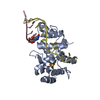
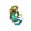
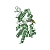
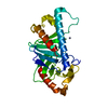
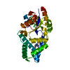
| Title | Perdeuterated mouse CNPase catalytic domain at atomic resolution | |||||||||
 Components Components | 2', 3'-CYCLIC-NUCLEOTIDE 3'-PHOSPHODIESTERASE | |||||||||
 Keywords Keywords | HYDROLASE | |||||||||
| Function / homology |  Function and homology information Function and homology informationcyclic nucleotide catabolic process / 2',3'-cyclic-nucleotide 3'-phosphodiesterase / 2',3'-cyclic-nucleotide 3'-phosphodiesterase activity / oligodendrocyte differentiation / axonogenesis / adult locomotory behavior / response to toxic substance / melanosome / myelin sheath / extracellular space ...cyclic nucleotide catabolic process / 2',3'-cyclic-nucleotide 3'-phosphodiesterase / 2',3'-cyclic-nucleotide 3'-phosphodiesterase activity / oligodendrocyte differentiation / axonogenesis / adult locomotory behavior / response to toxic substance / melanosome / myelin sheath / extracellular space / RNA binding / membrane / cytoplasm Similarity search - Function | |||||||||
| Biological species |  | |||||||||
| Method |  X-RAY DIFFRACTION / SYNCHROTRON / MOLECULAR REPLACEMENT / Resolution: 1.04 Å X-RAY DIFFRACTION / SYNCHROTRON / MOLECULAR REPLACEMENT / Resolution: 1.04 Å | |||||||||
 Authors Authors | Laulumaa, S. / Kursula, P. | |||||||||
 Citation Citation | Journal: Sci Rep / Year: 2015 Title: Determinants of ligand binding and catalytic activity in the myelin enzyme 2',3'-cyclic nucleotide 3'-phosphodiesterase. Authors: Raasakka, A. / Myllykoski, M. / Laulumaa, S. / Lehtimaki, M. / Hartlein, M. / Moulin, M. / Kursula, I. / Kursula, P. | |||||||||
| History |
|
- Structure visualization
| Structure viewer | Molecule: MolmilJmol/JSmol |
|---|
- Downloads & links
-Download
| PDBx/mmCIF format | 5ae0.cif.gz | 174.3 KB | Display | PDBx/mmCIF format |
|---|---|---|---|---|
| PDB format | pdb5ae0.ent.gz | 143.7 KB | Display | PDB format |
| PDBx/mmJSON format | 5ae0.json.gz | Tree view | PDBx/mmJSON format | |
| Others |  Other downloads Other downloads |
-Validation report
| Arichive directory | https://data.pdbj.org/pub/pdb/validation_reports/ae/5ae0ftp://data.pdbj.org/pub/pdb/validation_reports/ae/5ae0 | HTTPS FTP |
|---|
-Related structure data
| Related structure data |  4wbiC  4wblC  4wc9C  4wcaC  4wcbC  4wccC  4wdaC  4wdbC  4wddC  4wdeC  4wdfC  4wdgC  4wdhC  4wexC  4wfrC  2wutS S: Starting model for refinement C: citing same article ( |
|---|---|
| Similar structure data |


-Links
 PDBj
PDBj- Assembly
| Deposited unit | 
| ||||||||
|---|---|---|---|---|---|---|---|---|---|
| 1 |
| ||||||||
| Unit cell |
|
-Components
| #1: Protein | Mass: 24293.928 Da / Num. of mol.: 1 / Fragment: CATALYTIC DOMAIN, UNP RESIDUES 179-398 Source method: isolated from a genetically manipulated source Details: PERDEUTERATED / Source: (gene. exp.)  References: UniProt: P16330, 2',3'-cyclic-nucleotide 3'-phosphodiesterase |
|---|---|
| #2: Chemical | ChemComp-CIT /   Mass: 192.124 Da / Num. of mol.: 1 / Source method: obtained synthetically / Formula: C6H8O7 Mass: 192.124 Da / Num. of mol.: 1 / Source method: obtained synthetically / Formula: C6H8O7 |
| #3: Chemical | ChemComp-CL /   Mass: 35.453 Da / Num. of mol.: 1 / Source method: obtained synthetically / Formula: Cl Mass: 35.453 Da / Num. of mol.: 1 / Source method: obtained synthetically / Formula: Cl |
| #4: Water | ChemComp-HOH /  Mass: 18.015 Da / Num. of mol.: 282 / Source method: isolated from a natural source / Formula: H2O Mass: 18.015 Da / Num. of mol.: 282 / Source method: isolated from a natural source / Formula: H2O |
-Experimental details
-Experiment
| Experiment | Method: X-RAY DIFFRACTION / Number of used crystals: 1 |
|---|
- Sample preparation
| Crystal | Density Matthews: 1.97 Å3/Da / Density % sol: 37.59 % / Description: NONE |
|---|---|
| Crystal grow | Method: vapor diffusion, sitting drop / pH: 3.5 / Details: 25% PEG3350, 50 MM ACETATE, PD 3.5 |
-Data collection
| Diffraction | Mean temperature: 100 K |
|---|---|
| Diffraction source | Source: SYNCHROTRON / Site: MAX II  / Beamline: I911-3 / Wavelength: 0.913 / Beamline: I911-3 / Wavelength: 0.913 |
| Detector | Type: MARMOSAIC 225 mm CCD / Detector: CCD / Date: Jan 24, 2013 |
| Radiation | Protocol: SINGLE WAVELENGTH / Monochromatic (M) / Laue (L): M / Scattering type: x-ray |
| Radiation wavelength | Wavelength: 0.913 Å / Relative weight: 1 |
| Reflection | Resolution: 1.04→30 Å / Num. obs: 90347 / % possible obs: 99.7 % / Observed criterion σ(I): -3 / Redundancy: 3.7 % / Biso Wilson estimate: 8.77 Å2 / Rmerge(I) obs: 0.07 / Net I/σ(I): 9.6 |
| Reflection shell | Resolution: 1.04→1.07 Å / Redundancy: 2.9 % / Rmerge(I) obs: 1.26 / Mean I/σ(I) obs: 1.1 / % possible all: 99.1 |
- Processing
| Software |
| ||||||||||||||||||||||||||||||||||||||||||||||||||||||||||||||||||||||||||||||||||||||||||||||||||
|---|---|---|---|---|---|---|---|---|---|---|---|---|---|---|---|---|---|---|---|---|---|---|---|---|---|---|---|---|---|---|---|---|---|---|---|---|---|---|---|---|---|---|---|---|---|---|---|---|---|---|---|---|---|---|---|---|---|---|---|---|---|---|---|---|---|---|---|---|---|---|---|---|---|---|---|---|---|---|---|---|---|---|---|---|---|---|---|---|---|---|---|---|---|---|---|---|---|---|---|
| Refinement | Method to determine structure: MOLECULAR REPLACEMENT Starting model: PDB ENTRY 2WUT Resolution: 1.04→29.287 Å / SU ML: 0.1 / σ(F): 1.99 / Phase error: 17.06 / Stereochemistry target values: ML
| ||||||||||||||||||||||||||||||||||||||||||||||||||||||||||||||||||||||||||||||||||||||||||||||||||
| Solvent computation | Shrinkage radii: 0.9 Å / VDW probe radii: 1.11 Å / Solvent model: FLAT BULK SOLVENT MODEL | ||||||||||||||||||||||||||||||||||||||||||||||||||||||||||||||||||||||||||||||||||||||||||||||||||
| Refinement step | Cycle: LAST / Resolution: 1.04→29.287 Å
| ||||||||||||||||||||||||||||||||||||||||||||||||||||||||||||||||||||||||||||||||||||||||||||||||||
| Refine LS restraints |
| ||||||||||||||||||||||||||||||||||||||||||||||||||||||||||||||||||||||||||||||||||||||||||||||||||
| LS refinement shell |
|
