 Movie
Movie Controller

[English] 日本語
 Yorodumi
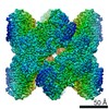
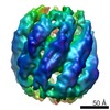
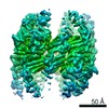
- PDB-4xci: Crystal structure of a hexadecameric TF55 complex from S. solfata... -
+ Open data

- Basic information
| Entry | Database: PDB / ID: 4xci | ||||||
|---|---|---|---|---|---|---|---|
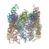
| Title | Crystal structure of a hexadecameric TF55 complex from S. solfataricus, crystal form II | ||||||
 Components Components |
| ||||||
 Keywords Keywords | CHAPERONE / Protein Folding / Thermosomes / Chaperonin | ||||||
| Function / homology |  Function and homology information Function and homology informationATP-dependent protein folding chaperone / unfolded protein binding / protein folding / ATP hydrolysis activity / ATP binding / identical protein binding Similarity search - Function | ||||||
| Biological species |   Sulfolobus solfataricus (archaea) Sulfolobus solfataricus (archaea) | ||||||
| Method |  X-RAY DIFFRACTION / SYNCHROTRON / MOLECULAR REPLACEMENT / Resolution: 3.0023 Å X-RAY DIFFRACTION / SYNCHROTRON / MOLECULAR REPLACEMENT / Resolution: 3.0023 Å | ||||||
 Authors Authors | Stewart, A.G. / Chaston, J.J. / Smits, C. / Stock, D. | ||||||
| Funding support |  Australia, 1items Australia, 1items
| ||||||
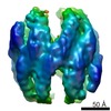
 Citation Citation | Journal: Structure / Year: 2016 Title: Structural and Functional Insights into the Evolution and Stress Adaptation of Type II Chaperonins. Authors: Jessica J Chaston / Callum Smits / David Aragão / Andrew S W Wong / Bilal Ahsan / Sara Sandin / Sudheer K Molugu / Sanjay K Molugu / Ricardo A Bernal / Daniela Stock / Alastair G Stewart /   Abstract: Chaperonins are essential biological complexes assisting protein folding in all kingdoms of life. Whereas homooligomeric bacterial GroEL binds hydrophobic substrates non-specifically, the ...Chaperonins are essential biological complexes assisting protein folding in all kingdoms of life. Whereas homooligomeric bacterial GroEL binds hydrophobic substrates non-specifically, the heterooligomeric eukaryotic CCT binds specifically to distinct classes of substrates. Sulfolobales, which survive in a wide range of temperatures, have evolved three different chaperonin subunits (α, β, γ) that form three distinct complexes tailored for different substrate classes at cold, normal, and elevated temperatures. The larger octadecameric β complexes cater for substrates under heat stress, whereas smaller hexadecameric αβ complexes prevail under normal conditions. The cold-shock complex contains all three subunits, consistent with greater substrate specificity. Structural analysis using crystallography and electron microscopy reveals the geometry of these complexes and shows a novel arrangement of the α and β subunits in the hexadecamer enabling incorporation of the γ subunit. | ||||||
| History |
|
- Structure visualization
| Structure viewer | Molecule: MolmilJmol/JSmol |
|---|
- Downloads & links
-Download
| PDBx/mmCIF format | 4xci.cif.gz | 289.2 KB | Display | PDBx/mmCIF format |
|---|---|---|---|---|
| PDB format | pdb4xci.ent.gz | 235.2 KB | Display | PDB format |
| PDBx/mmJSON format | 4xci.json.gz | Tree view | PDBx/mmJSON format | |
| Others |  Other downloads Other downloads |
-Validation report
| Summary document | 4xci_validation.pdf.gz | 441.7 KB | Display | wwPDB validaton report |
|---|---|---|---|---|
| Full document | 4xci_full_validation.pdf.gz | 449.3 KB | Display | |
| Data in XML | 4xci_validation.xml.gz | 26.6 KB | Display | |
| Data in CIF | 4xci_validation.cif.gz | 36 KB | Display | |
| Arichive directory | https://data.pdbj.org/pub/pdb/validation_reports/xc/4xciftp://data.pdbj.org/pub/pdb/validation_reports/xc/4xci | HTTPS FTP |
-Related structure data
| Related structure data | 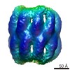 6291C 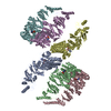 4xcdC  4xcgC 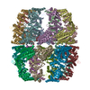 3j1bS 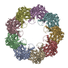 3ko1S S: Starting model for refinement C: citing same article ( |
|---|---|
| Similar structure data |
-Links
 PDBj
PDBj
- Assembly
| Deposited unit | 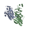
| ||||||||
|---|---|---|---|---|---|---|---|---|---|
| 1 | x 8
| ||||||||
| Unit cell |
| ||||||||
| Symmetry | Point symmetry: (Schoenflies symbol: D4 (2x4 fold dihedral)) | ||||||||
| Details | Electron Microscopy and MALLS confirm size and stoichiometry of a hexadecameric complex |
-Components
| #1: Protein | Mass: 60453.465 Da / Num. of mol.: 1 / Source method: isolated from a natural source Source: (natural) Sulfolobus solfataricus (strain ATCC 35092 / DSM 1617 / JCM 11322 / P2) (archaea)Strain: ATCC 35092 / DSM 1617 / JCM 11322 / P2 / References: UniProt: Q9V2T8 |
|---|---|
| #2: Protein | Mass: 59754.027 Da / Num. of mol.: 1 / Source method: isolated from a natural source Source: (natural) Sulfolobus solfataricus (strain ATCC 35092 / DSM 1617 / JCM 11322 / P2) (archaea)Strain: ATCC 35092 / DSM 1617 / JCM 11322 / P2 / References: UniProt: Q9V2S9 |
-Experimental details
-Experiment
| Experiment | Method: X-RAY DIFFRACTION / Number of used crystals: 1 |
|---|
- Sample preparation
| Crystal | Density Matthews: 3.49 Å3/Da / Density % sol: 64.75 % |
|---|---|
| Crystal grow | Temperature: 293 K / Method: vapor diffusion, hanging drop / pH: 7.9 Details: Tris-HCl, PEG 2000, 2-propanol, strontium chloride, TMAO |
-Data collection
| Diffraction | Mean temperature: 100 K | |||||||||||||||||||||||||||
|---|---|---|---|---|---|---|---|---|---|---|---|---|---|---|---|---|---|---|---|---|---|---|---|---|---|---|---|---|
| Diffraction source | Source: SYNCHROTRON / Site: Australian Synchrotron / Beamline: MX2 / Wavelength: 0.9537 Å | |||||||||||||||||||||||||||
| Detector | Type: ADSC QUANTUM 315r / Detector: CCD / Date: Jul 11, 2013 | |||||||||||||||||||||||||||
| Radiation | Protocol: SINGLE WAVELENGTH / Monochromatic (M) / Laue (L): M / Scattering type: x-ray | |||||||||||||||||||||||||||
| Radiation wavelength | Wavelength: 0.9537 Å / Relative weight: 1 | |||||||||||||||||||||||||||
| Reflection | Resolution: 3→48.83 Å / Num. obs: 34477 / % possible obs: 100 % / Redundancy: 14.7 % / Biso Wilson estimate: 70.99 Å2 / CC1/2: 0.997 / Rmerge(I) obs: 0.381 / Rpim(I) all: 0.102 / Net I/σ(I): 6.8 / Num. measured all: 507888 / Scaling rejects: 128 | |||||||||||||||||||||||||||
| Reflection shell | Diffraction-ID: 1 / Rejects: _
|
- Processing
| Software |
| ||||||||||||||||||||||||||||||||||||||||||||||||||||||||||||||||||||||
|---|---|---|---|---|---|---|---|---|---|---|---|---|---|---|---|---|---|---|---|---|---|---|---|---|---|---|---|---|---|---|---|---|---|---|---|---|---|---|---|---|---|---|---|---|---|---|---|---|---|---|---|---|---|---|---|---|---|---|---|---|---|---|---|---|---|---|---|---|---|---|---|
| Refinement | Method to determine structure: MOLECULAR REPLACEMENT Starting model: 3KO1, 3J1B Resolution: 3.0023→41.907 Å / SU ML: 0.38 / Cross valid method: FREE R-VALUE / σ(F): 1.35 / Phase error: 30.28 / Stereochemistry target values: ML
| ||||||||||||||||||||||||||||||||||||||||||||||||||||||||||||||||||||||
| Solvent computation | Shrinkage radii: 0.9 Å / VDW probe radii: 1.11 Å / Solvent model: FLAT BULK SOLVENT MODEL | ||||||||||||||||||||||||||||||||||||||||||||||||||||||||||||||||||||||
| Displacement parameters | Biso max: 327.01 Å2 / Biso mean: 91.5051 Å2 / Biso min: 12.83 Å2 | ||||||||||||||||||||||||||||||||||||||||||||||||||||||||||||||||||||||
| Refinement step | Cycle: final / Resolution: 3.0023→41.907 Å
| ||||||||||||||||||||||||||||||||||||||||||||||||||||||||||||||||||||||
| Refine LS restraints |
| ||||||||||||||||||||||||||||||||||||||||||||||||||||||||||||||||||||||
| LS refinement shell | Refine-ID: X-RAY DIFFRACTION / Total num. of bins used: 9
|
