 Movie
Movie Controller

[English] 日本語
 Yorodumi
- PDB-4uv6: Crystal structure of apical membrane antigen 1 from Plasmodium kn... -
+ Open data

- Basic information
| Entry | Database: PDB / ID: 4uv6 | ||||||
|---|---|---|---|---|---|---|---|
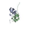
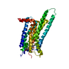
| Title | Crystal structure of apical membrane antigen 1 from Plasmodium knowlesi | ||||||
 Components Components | APICAL MEROZOITE ANTIGEN 1 | ||||||
 Keywords Keywords | CELL INVASION / MALARIA / VACCINE CANDIDATE | ||||||
| Function / homology |  Function and homology information Function and homology information | ||||||
| Biological species |  | ||||||
| Method |  X-RAY DIFFRACTION / SYNCHROTRON / MOLECULAR REPLACEMENT / Resolution: 2.45 Å X-RAY DIFFRACTION / SYNCHROTRON / MOLECULAR REPLACEMENT / Resolution: 2.45 Å | ||||||
 Authors Authors | Vulliez-Le Normand, B. / Saul, F.A. / Bentley, G.A. | ||||||
 Citation Citation | Journal: Plos One / Year: 2015 Title: Crystal Structure of Plasmodium Knowlesi Apical Membrane Antigen 1 and its Complex with an Invasion-Inhibitory Monoclonal Antibody. Authors: Vulliez-Le Normand, B. / Faber, B.W. / Saul, F.A. / Van Der Eijk, M. / Thomas, A.W. / Singh, B. / Kocken, C.H.M. / Bentley, G.A. | ||||||
| History |
|
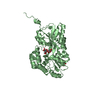
- Structure visualization
| Structure viewer | Molecule: MolmilJmol/JSmol |
|---|
- Downloads & links
-Download
| PDBx/mmCIF format | 4uv6.cif.gz | 155 KB | Display | PDBx/mmCIF format |
|---|---|---|---|---|
| PDB format | pdb4uv6.ent.gz | 122.6 KB | Display | PDB format |
| PDBx/mmJSON format | 4uv6.json.gz | Tree view | PDBx/mmJSON format | |
| Others |  Other downloads Other downloads |
-Validation report
| Arichive directory | https://data.pdbj.org/pub/pdb/validation_reports/uv/4uv6ftp://data.pdbj.org/pub/pdb/validation_reports/uv/4uv6 | HTTPS FTP |
|---|
-Related structure data
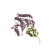
| Related structure data |  4uaoC  1w81S S: Starting model for refinement C: citing same article ( |
|---|---|
| Similar structure data |


-Links
 PDBj
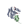
PDBj- Assembly
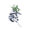
| Deposited unit | 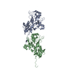
| ||||||||
|---|---|---|---|---|---|---|---|---|---|
| 1 | 
| ||||||||
| 2 | 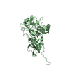
| ||||||||
| Unit cell |
| ||||||||
| Noncrystallographic symmetry (NCS) | NCS oper: (Code: given Matrix: (-0.95306, 0.04609, -0.29926), Vector: |
-Components
| #1: Protein | Mass: 42511.449 Da / Num. of mol.: 2 / Fragment: UNP RESIDUES 43-387 / Mutation: YES Source method: isolated from a genetically manipulated source Source: (gene. exp.)  PICHIA PASTORIS (fungus) / Strain (production host): KM71H / References: UniProt: B3L5E1, UniProt: A0A384KGX8*PLUS PICHIA PASTORIS (fungus) / Strain (production host): KM71H / References: UniProt: B3L5E1, UniProt: A0A384KGX8*PLUS#2: Water | ChemComp-HOH / |  Mass: 18.015 Da / Num. of mol.: 386 / Source method: isolated from a natural source / Formula: H2O Mass: 18.015 Da / Num. of mol.: 386 / Source method: isolated from a natural source / Formula: H2OHas protein modification | Y | |
|---|
-Experimental details
-Experiment
| Experiment | Method: X-RAY DIFFRACTION / Number of used crystals: 1 |
|---|
- Sample preparation
| Crystal | Density Matthews: 2.93 Å3/Da / Density % sol: 58.07 % / Description: NONE |
|---|---|
| Crystal grow | pH: 8.4 / Details: 0.86M SODIUM CITRATE, 0.1M HEPES PH 8.4 |
-Data collection
| Diffraction | Mean temperature: 100 K |
|---|---|
| Diffraction source | Source: SYNCHROTRON / Site: ESRF  / Beamline: ID14-4 / Wavelength: 0.976 / Beamline: ID14-4 / Wavelength: 0.976 |
| Detector | Type: ADSC CCD / Detector: CCD / Date: Nov 5, 2007 |
| Radiation | Protocol: SINGLE WAVELENGTH / Monochromatic (M) / Laue (L): M / Scattering type: x-ray |
| Radiation wavelength | Wavelength: 0.976 Å / Relative weight: 1 |
| Reflection | Resolution: 2.45→47.09 Å / Num. obs: 34848 / % possible obs: 97.3 % / Observed criterion σ(I): -3 / Redundancy: 2.9 % / Biso Wilson estimate: 42.45 Å2 / Rmerge(I) obs: 0.1 / Net I/σ(I): 9.7 |
| Reflection shell | Resolution: 2.45→2.58 Å / Redundancy: 2.5 % / Rmerge(I) obs: 0.5 / Mean I/σ(I) obs: 1.9 / % possible all: 93.3 |
- Processing
| Software |
| ||||||||||||||||||||||||||||||||||||||||||||||||||||||||||||||||||||||||||||||||||||||||||||||||||||||||||||||||||
|---|---|---|---|---|---|---|---|---|---|---|---|---|---|---|---|---|---|---|---|---|---|---|---|---|---|---|---|---|---|---|---|---|---|---|---|---|---|---|---|---|---|---|---|---|---|---|---|---|---|---|---|---|---|---|---|---|---|---|---|---|---|---|---|---|---|---|---|---|---|---|---|---|---|---|---|---|---|---|---|---|---|---|---|---|---|---|---|---|---|---|---|---|---|---|---|---|---|---|---|---|---|---|---|---|---|---|---|---|---|---|---|---|---|---|---|
| Refinement | Method to determine structure: MOLECULAR REPLACEMENT Starting model: PDB ENTRY 1W81 Resolution: 2.45→47.09 Å / Cor.coef. Fo:Fc: 0.9181 / Cor.coef. Fo:Fc free: 0.8668 / SU R Cruickshank DPI: 0.299 / Cross valid method: THROUGHOUT / σ(F): 0 / SU R Blow DPI: 0.307 / SU Rfree Blow DPI: 0.228 / SU Rfree Cruickshank DPI: 0.228
| ||||||||||||||||||||||||||||||||||||||||||||||||||||||||||||||||||||||||||||||||||||||||||||||||||||||||||||||||||
| Displacement parameters | Biso mean: 45.48 Å2
| ||||||||||||||||||||||||||||||||||||||||||||||||||||||||||||||||||||||||||||||||||||||||||||||||||||||||||||||||||
| Refine analyze | Luzzati coordinate error obs: 0.281 Å | ||||||||||||||||||||||||||||||||||||||||||||||||||||||||||||||||||||||||||||||||||||||||||||||||||||||||||||||||||
| Refinement step | Cycle: LAST / Resolution: 2.45→47.09 Å
| ||||||||||||||||||||||||||||||||||||||||||||||||||||||||||||||||||||||||||||||||||||||||||||||||||||||||||||||||||
| Refine LS restraints |
| ||||||||||||||||||||||||||||||||||||||||||||||||||||||||||||||||||||||||||||||||||||||||||||||||||||||||||||||||||
| LS refinement shell | Resolution: 2.45→2.52 Å / Total num. of bins used: 17
|
