 Movie
Movie Controller

[English] 日本語
 Yorodumi
- PDB-5b3w: Crystal structure of hPin1 WW domain (5-15) fused with maltose-bi... -
+ Open data

- Basic information
| Entry | Database: PDB / ID: 5b3w | |||||||||
|---|---|---|---|---|---|---|---|---|---|---|
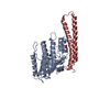
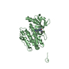
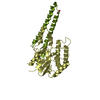
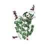
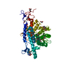
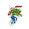
| Title | Crystal structure of hPin1 WW domain (5-15) fused with maltose-binding protein in C2221 form | |||||||||
 Components Components | Peptidyl-prolyl cis-trans isomerase NIMA-interacting 1,Maltose-binding periplasmic protein | |||||||||
 Keywords Keywords | ISOMERASE / SUGAR BINDING PROTEIN | |||||||||
| Function / homology |  Function and homology information Function and homology informationcis-trans isomerase activity / phosphothreonine residue binding / negative regulation of cell motility / regulation of protein localization to nucleus / negative regulation of brown fat cell differentiation / mitogen-activated protein kinase kinase binding / ubiquitin ligase activator activity / GTPase activating protein binding / : / protein peptidyl-prolyl isomerization ...cis-trans isomerase activity / phosphothreonine residue binding / negative regulation of cell motility / regulation of protein localization to nucleus / negative regulation of brown fat cell differentiation / mitogen-activated protein kinase kinase binding / ubiquitin ligase activator activity / GTPase activating protein binding / : / protein peptidyl-prolyl isomerization / regulation of mitotic nuclear division / negative regulation of SMAD protein signal transduction / detection of maltose stimulus / PI5P Regulates TP53 Acetylation / negative regulation of amyloid-beta formation / maltose transport complex / carbohydrate transport / cytoskeletal motor activity / phosphoserine residue binding / RHO GTPases Activate NADPH Oxidases / carbohydrate transmembrane transporter activity / maltose binding / maltose transport / maltodextrin transmembrane transport / postsynaptic cytosol / Rho protein signal transduction / ATP-binding cassette (ABC) transporter complex, substrate-binding subunit-containing / ATP-binding cassette (ABC) transporter complex / regulation of cytokinesis / peptidylprolyl isomerase / peptidyl-prolyl cis-trans isomerase activity / cell chemotaxis / Negative regulators of DDX58/IFIH1 signaling / negative regulation of transforming growth factor beta receptor signaling pathway / regulation of protein stability / phosphoprotein binding / negative regulation of ERK1 and ERK2 cascade / negative regulation of protein catabolic process / positive regulation of protein phosphorylation / synapse organization / beta-catenin binding / protein destabilization / tau protein binding / ISG15 antiviral mechanism / neuron differentiation / positive regulation of canonical Wnt signaling pathway / outer membrane-bounded periplasmic space / regulation of gene expression / midbody / cellular response to hypoxia / Regulation of TP53 Activity through Phosphorylation / response to hypoxia / periplasmic space / nuclear speck / ciliary basal body / protein stabilization / DNA damage response / glutamatergic synapse / positive regulation of transcription by RNA polymerase II / nucleoplasm / membrane / nucleus / cytosol / cytoplasm Similarity search - Function | |||||||||
| Biological species |  Homo sapiens (human) Homo sapiens (human) | |||||||||
| Method |  X-RAY DIFFRACTION / SYNCHROTRON / MOLECULAR REPLACEMENT / Resolution: 2.4 Å X-RAY DIFFRACTION / SYNCHROTRON / MOLECULAR REPLACEMENT / Resolution: 2.4 Å | |||||||||
 Authors Authors | Hanazono, Y. / Takeda, K. / Miki, K. | |||||||||
 Citation Citation | Journal: Sci Rep / Year: 2016 Title: Structural studies of the N-terminal fragments of the WW domain: Insights into co-translational folding of a beta-sheet protein Authors: Hanazono, Y. / Takeda, K. / Miki, K. | |||||||||
| History |
|
- Structure visualization
| Structure viewer | Molecule: MolmilJmol/JSmol |
|---|
- Downloads & links
-Download
| PDBx/mmCIF format | 5b3w.cif.gz | 173.6 KB | Display | PDBx/mmCIF format |
|---|---|---|---|---|
| PDB format | pdb5b3w.ent.gz | 136 KB | Display | PDB format |
| PDBx/mmJSON format | 5b3w.json.gz | Tree view | PDBx/mmJSON format | |
| Others |  Other downloads Other downloads |
-Validation report
| Arichive directory | https://data.pdbj.org/pub/pdb/validation_reports/b3/5b3wftp://data.pdbj.org/pub/pdb/validation_reports/b3/5b3w | HTTPS FTP |
|---|
-Related structure data
| Related structure data | 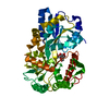 5b3xC 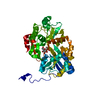 5b3yC 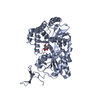 5b3zC 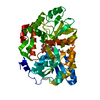 5bmyC  1anfS C: citing same article ( S: Starting model for refinement |
|---|---|
| Similar structure data |
-Links
 PDBj
PDBj


- Assembly
| Deposited unit | 
| ||||||||
|---|---|---|---|---|---|---|---|---|---|
| 1 | 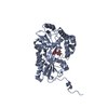
| ||||||||
| 2 | 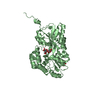
| ||||||||
| Unit cell |
|
-Components
| #1: Protein | Mass: 42054.609 Da / Num. of mol.: 2 Fragment: UNP(Q13526) residues 5-15,UNP(P0AEX9) residues 27-393 Mutation: R382N Source method: isolated from a genetically manipulated source Source: (gene. exp.) Homo sapiens (human), (gene. exp.) Gene: PIN1, malE / Strain: K-12 / Production host: #2: Polysaccharide |   Source method: isolated from a genetically manipulated source Details: oligosaccharide / References: alpha-maltose #3: Chemical | ChemComp-CIT / |   Mass: 192.124 Da / Num. of mol.: 1 / Source method: obtained synthetically / Formula: C6H8O7 Mass: 192.124 Da / Num. of mol.: 1 / Source method: obtained synthetically / Formula: C6H8O7#4: Water | ChemComp-HOH / |  Mass: 18.015 Da / Num. of mol.: 591 / Source method: isolated from a natural source / Formula: H2O Mass: 18.015 Da / Num. of mol.: 591 / Source method: isolated from a natural source / Formula: H2O |
|---|
-Experimental details
-Experiment
| Experiment | Method: X-RAY DIFFRACTION / Number of used crystals: 1 |
|---|
- Sample preparation
| Crystal | Density Matthews: 3.17 Å3/Da / Density % sol: 61.22 % |
|---|---|
| Crystal grow | Temperature: 293 K / Method: vapor diffusion, sitting drop / Details: 2.2 M DL-malic acid |
-Data collection
| Diffraction | Mean temperature: 100 K |
|---|---|
| Diffraction source | Source: SYNCHROTRON / Site: SPring-8  / Beamline: BL41XU / Wavelength: 1 Å / Beamline: BL41XU / Wavelength: 1 Å |
| Detector | Type: RAYONIX MX225HE / Detector: CCD / Date: Jun 27, 2012 |
| Radiation | Protocol: SINGLE WAVELENGTH / Monochromatic (M) / Laue (L): M / Scattering type: x-ray |
| Radiation wavelength | Wavelength: 1 Å / Relative weight: 1 |
| Reflection | Resolution: 2.4→50 Å / Num. obs: 41976 / % possible obs: 99.5 % / Redundancy: 6.3 % / Rsym value: 0.131 / Net I/σ(I): 14.4 |
| Reflection shell | Resolution: 2.4→2.44 Å / Mean I/σ(I) obs: 3.7 / % possible all: 99.1 |
- Processing
| Software |
| ||||||||||||||||||||||||||||||||||||||||||||||||||||||||||||||||||||||||||||||||||||||||||||||||||||||||||||||||
|---|---|---|---|---|---|---|---|---|---|---|---|---|---|---|---|---|---|---|---|---|---|---|---|---|---|---|---|---|---|---|---|---|---|---|---|---|---|---|---|---|---|---|---|---|---|---|---|---|---|---|---|---|---|---|---|---|---|---|---|---|---|---|---|---|---|---|---|---|---|---|---|---|---|---|---|---|---|---|---|---|---|---|---|---|---|---|---|---|---|---|---|---|---|---|---|---|---|---|---|---|---|---|---|---|---|---|---|---|---|---|---|---|---|
| Refinement | Method to determine structure: MOLECULAR REPLACEMENT Starting model: 1ANF Resolution: 2.4→38.573 Å / SU ML: 0.26 / Cross valid method: FREE R-VALUE / σ(F): 1.35 / Phase error: 22.83
| ||||||||||||||||||||||||||||||||||||||||||||||||||||||||||||||||||||||||||||||||||||||||||||||||||||||||||||||||
| Solvent computation | Shrinkage radii: 0.9 Å / VDW probe radii: 1.11 Å | ||||||||||||||||||||||||||||||||||||||||||||||||||||||||||||||||||||||||||||||||||||||||||||||||||||||||||||||||
| Refinement step | Cycle: LAST / Resolution: 2.4→38.573 Å
| ||||||||||||||||||||||||||||||||||||||||||||||||||||||||||||||||||||||||||||||||||||||||||||||||||||||||||||||||
| Refine LS restraints |
| ||||||||||||||||||||||||||||||||||||||||||||||||||||||||||||||||||||||||||||||||||||||||||||||||||||||||||||||||
| LS refinement shell |
|
