 Movie
Movie Controller


+ Open data

- Basic information
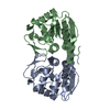
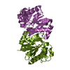
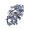
| Entry | Database: PDB / ID: 4s0z | ||||||
|---|---|---|---|---|---|---|---|
| Title | Crystal structure of M26V human DJ-1 | ||||||
 Components Components | Protein DJ-1 | ||||||
 Keywords Keywords |  CHAPERONE / DJ-1/ThiJ/PfpI Superfamily / Oxidative stress response CHAPERONE / DJ-1/ThiJ/PfpI Superfamily / Oxidative stress response | ||||||
| Function / homology |  Function and homology information Function and homology informationtyrosine 3-monooxygenase activator activity / cellular response to glyoxal / L-dopa decarboxylase activator activity / peptidyl-cysteine deglycation / peptidyl-arginine deglycation / peptidyl-lysine deglycation / protein deglycation, methylglyoxal removal / glutathione deglycation / detoxification of hydrogen peroxide / methylglyoxal catabolic process to lactate ...tyrosine 3-monooxygenase activator activity / cellular response to glyoxal / L-dopa decarboxylase activator activity / peptidyl-cysteine deglycation / peptidyl-arginine deglycation / peptidyl-lysine deglycation / protein deglycation, methylglyoxal removal / glutathione deglycation / detoxification of hydrogen peroxide / methylglyoxal catabolic process to lactate / guanine deglycation, methylglyoxal removal / cellular detoxification of methylglyoxal / regulation of supramolecular fiber organization / negative regulation of death-inducing signaling complex assembly / negative regulation of TRAIL-activated apoptotic signaling pathway / positive regulation of pyrroline-5-carboxylate reductase activity / positive regulation of tyrosine 3-monooxygenase activity / positive regulation of L-dopa biosynthetic process / positive regulation of L-dopa decarboxylase activity / negative regulation of hydrogen peroxide-induced neuron intrinsic apoptotic signaling pathway / glyoxalase (glycolic acid-forming) activity / negative regulation of protein K48-linked deubiquitination / negative regulation of ubiquitin-specific protease activity / protein deglycation, glyoxal removal / glycolate biosynthetic process / guanine deglycation, glyoxal removal / glyoxal metabolic process / negative regulation of nitrosative stress-induced intrinsic apoptotic signaling pathway / detection of oxidative stress / guanine deglycation / detoxification of mercury ion / positive regulation of mitochondrial electron transport, NADH to ubiquinone / protein deglycase / methylglyoxal metabolic process / mercury ion binding / oxidoreductase activity, acting on peroxide as acceptor / protein deglycase activity / positive regulation of acute inflammatory response to antigenic stimulus / positive regulation of dopamine biosynthetic process / superoxide dismutase copper chaperone activity / positive regulation of NAD(P)H oxidase activity / positive regulation of autophagy of mitochondrion / lactate biosynthetic process / negative regulation of cysteine-type endopeptidase activity involved in apoptotic signaling pathway / cellular detoxification of aldehyde / positive regulation of superoxide dismutase activity / small protein activating enzyme binding / Hydrolases; Acting on ester bonds; Thioester hydrolases / regulation of oxidative stress-induced neuron intrinsic apoptotic signaling pathway / negative regulation of ubiquitin-protein transferase activity / peroxiredoxin activity / detoxification of copper ion / negative regulation of protein acetylation / negative regulation of oxidative stress-induced neuron intrinsic apoptotic signaling pathway / positive regulation of transcription regulatory region DNA binding / positive regulation of oxidative stress-induced intrinsic apoptotic signaling pathway / positive regulation of androgen receptor activity / membrane hyperpolarization / protein deglycosylation / negative regulation of protein sumoylation / oxygen sensor activity / regulation of androgen receptor signaling pathway / negative regulation of protein export from nucleus / negative regulation of intrinsic apoptotic signaling pathway in response to hydrogen peroxide / cupric ion binding / ubiquitin-like protein conjugating enzyme binding / insulin secretion / Hydrolases; Acting on carbon-nitrogen bonds, other than peptide bonds; In linear amides / dopamine uptake involved in synaptic transmission / positive regulation of reactive oxygen species biosynthetic process / nuclear androgen receptor binding / hydrogen peroxide metabolic process / ubiquitin-specific protease binding / cytokine binding / cuprous ion binding / single fertilization / membrane depolarization / negative regulation of proteasomal ubiquitin-dependent protein catabolic process / negative regulation of endoplasmic reticulum stress-induced intrinsic apoptotic signaling pathway / regulation of neuron apoptotic process / negative regulation of oxidative stress-induced intrinsic apoptotic signaling pathway / negative regulation of reactive oxygen species biosynthetic process / negative regulation of protein ubiquitination / activation of protein kinase B activity / mitochondrion organization / adult locomotory behavior / SUMOylation of transcription cofactors / regulation of mitochondrial membrane potential / negative regulation of protein phosphorylation / negative regulation of protein binding / positive regulation of interleukin-8 production / positive regulation of protein-containing complex assembly / negative regulation of extrinsic apoptotic signaling pathway / adherens junction / Late endosomal microautophagy / negative regulation of protein kinase activity / mitochondrial intermembrane space / PML body / cellular response to hydrogen peroxide / autophagySimilarity search - Function | ||||||
| Biological species |  Homo sapiens (human) Homo sapiens (human) | ||||||
| Method | X-RAY DIFFRACTION / MOLECULAR REPLACEMENT / Resolution: 1.45 Å | ||||||
 Authors Authors | Milkovic, N.M. / Wilson, M.A. | ||||||
 Citation Citation | Journal: Protein Sci. / Year: 2015 Title: Transient sampling of aggregation-prone conformations causes pathogenic instability of a parkinsonian mutant of DJ-1 at physiological temperature. Authors: Milkovic, N.M. / Catazaro, J. / Lin, J. / Halouska, S. / Kizziah, J.L. / Basiaga, S. / Cerny, R.L. / Powers, R. / Wilson, M.A. | ||||||
| History |
|
- Structure visualization
| Structure viewer | Molecule: MolmilJmol/JSmol |
|---|
- Downloads & links
-Download
| PDBx/mmCIF format | 4s0z.cif.gz | 102.5 KB | Display | PDBx/mmCIF format |
|---|---|---|---|---|
| PDB format | pdb4s0z.ent.gz | 78.9 KB | Display | PDB format |
| PDBx/mmJSON format | 4s0z.json.gz | Tree view | PDBx/mmJSON format | |
| Others |  Other downloads Other downloads |
-Validation report
| Arichive directory | https://data.pdbj.org/pub/pdb/validation_reports/s0/4s0zftp://data.pdbj.org/pub/pdb/validation_reports/s0/4s0z | HTTPS FTP |
|---|
-Related structure data
| Related structure data |  1p5fS S: Starting model for refinement |
|---|---|
| Similar structure data |


-Links
 PDBj
PDBj


- Assembly
| Deposited unit | 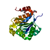
| ||||||||
|---|---|---|---|---|---|---|---|---|---|
| 1 | 
| ||||||||
| Unit cell |
|
-Components
| #1: Protein | Mass: 20167.262 Da / Num. of mol.: 1 / Mutation: M26V Source method: isolated from a genetically manipulated source Source: (gene. exp.) Homo sapiens (human) / Gene: PARK7 / Plasmid: pET15b / Production host:  Escherichia coli (E. coli) / Strain (production host): BL21(DE3) Escherichia coli (E. coli) / Strain (production host): BL21(DE3)References: UniProt: Q99497, Hydrolases; Acting on peptide bonds (peptidases) | ||||
|---|---|---|---|---|---|
| #2: Chemical | ChemComp-EDO / Ethylene glycol  Mass: 62.068 Da / Num. of mol.: 4 / Source method: obtained synthetically / Formula: C2H6O2 Mass: 62.068 Da / Num. of mol.: 4 / Source method: obtained synthetically / Formula: C2H6O2#3: Chemical | ChemComp-ACT / | Acetate  Mass: 59.044 Da / Num. of mol.: 1 / Source method: obtained synthetically / Formula: C2H3O2 Mass: 59.044 Da / Num. of mol.: 1 / Source method: obtained synthetically / Formula: C2H3O2#4: Water | ChemComp-HOH / | Water Mass: 18.015 Da / Num. of mol.: 276 / Source method: isolated from a natural source / Formula: H2O Mass: 18.015 Da / Num. of mol.: 276 / Source method: isolated from a natural source / Formula: H2O |
-Experimental details
-Experiment
| Experiment | Method: X-RAY DIFFRACTION / Number of used crystals: 1 |
|---|
- Sample preparation
| Crystal | Density Matthews: 3.03 Å3/Da / Density % sol: 59.45 % |
|---|---|
| Crystal grow | Temperature: 298 K / Method: vapor diffusion, sitting drop / pH: 9 Details: 100 mM Tris-HCl pH 9.0, 200 mM sodium acetate trihydrate, 25% PEG 4000 and 3 mM DTT, VAPOR DIFFUSION, SITTING DROP, temperature 298K |
-Data collection
| Diffraction | Mean temperature: 100 K |
|---|---|
| Diffraction source | Source: ROTATING ANODE / Type: RIGAKU MICROMAX-007 / Wavelength: 1.54 Å |
| Detector | Type: RIGAKU RAXIS IV++ / Detector: IMAGE PLATE / Date: Jun 26, 2014 / Details: Osmic blue confocal optics |
| Radiation | Monochromator: confocal / Protocol: SINGLE WAVELENGTH / Monochromatic (M) / Laue (L): M / Scattering type: x-ray |
| Radiation wavelength | Wavelength: 1.54 Å / Relative weight: 1 |
| Reflection | Resolution: 1.45→65 Å / Num. all: 43750 / Num. obs: 43750 / % possible obs: 99.7 % / Observed criterion σ(F): 0 / Observed criterion σ(I): 0 / Redundancy: 20.4 % / Rmerge(I) obs: 0.062 / Net I/σ(I): 56.6 |
| Reflection shell | Resolution: 1.45→1.5 Å / Redundancy: 18.2 % / Rmerge(I) obs: 0.523 / Mean I/σ(I) obs: 5 / % possible all: 97 |
- Processing
| Software |
| ||||||||||||||||||||||||||||||||||||||||||||||||||||||||||||||||||||||||||||||||||||||||||||||||||||||||||||||||||||||||||||||||||||||||||||||||||||||||||||||||||||||||||||||||||||||
|---|---|---|---|---|---|---|---|---|---|---|---|---|---|---|---|---|---|---|---|---|---|---|---|---|---|---|---|---|---|---|---|---|---|---|---|---|---|---|---|---|---|---|---|---|---|---|---|---|---|---|---|---|---|---|---|---|---|---|---|---|---|---|---|---|---|---|---|---|---|---|---|---|---|---|---|---|---|---|---|---|---|---|---|---|---|---|---|---|---|---|---|---|---|---|---|---|---|---|---|---|---|---|---|---|---|---|---|---|---|---|---|---|---|---|---|---|---|---|---|---|---|---|---|---|---|---|---|---|---|---|---|---|---|---|---|---|---|---|---|---|---|---|---|---|---|---|---|---|---|---|---|---|---|---|---|---|---|---|---|---|---|---|---|---|---|---|---|---|---|---|---|---|---|---|---|---|---|---|---|---|---|---|---|
| Refinement | Method to determine structure: MOLECULAR REPLACEMENT Starting model: 1P5F Resolution: 1.45→64.99 Å / Cor.coef. Fo:Fc: 0.982 / Cor.coef. Fo:Fc free: 0.978 / SU B: 1.199 / SU ML: 0.021 / Cross valid method: THROUGHOUT / ESU R: 0.044 / ESU R Free: 0.041 / Stereochemistry target values: MAXIMUM LIKELIHOOD / Details: HYDROGENS HAVE BEEN ADDED IN THE RIDING POSITIONS
| ||||||||||||||||||||||||||||||||||||||||||||||||||||||||||||||||||||||||||||||||||||||||||||||||||||||||||||||||||||||||||||||||||||||||||||||||||||||||||||||||||||||||||||||||||||||
| Solvent computation | Ion probe radii: 0.8 Å / Shrinkage radii: 0.8 Å / VDW probe radii: 1.2 Å / Solvent model: MASK | ||||||||||||||||||||||||||||||||||||||||||||||||||||||||||||||||||||||||||||||||||||||||||||||||||||||||||||||||||||||||||||||||||||||||||||||||||||||||||||||||||||||||||||||||||||||
| Displacement parameters | Biso mean: 20.241 Å2
| ||||||||||||||||||||||||||||||||||||||||||||||||||||||||||||||||||||||||||||||||||||||||||||||||||||||||||||||||||||||||||||||||||||||||||||||||||||||||||||||||||||||||||||||||||||||
| Refinement step | Cycle: LAST / Resolution: 1.45→64.99 Å
| ||||||||||||||||||||||||||||||||||||||||||||||||||||||||||||||||||||||||||||||||||||||||||||||||||||||||||||||||||||||||||||||||||||||||||||||||||||||||||||||||||||||||||||||||||||||
| Refine LS restraints |
|