 Movie
Movie Controller


+ Open data

- Basic information
| Entry | Database: PDB / ID: 400000000 | ||||||
|---|---|---|---|---|---|---|---|
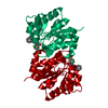
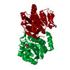
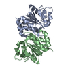
| Title | Crystal structure of Drosophila melanogaster DJ-1beta | ||||||
 Components Components | DJ-1 beta | ||||||
 Keywords Keywords | MOTOR PROTEIN / flavodoxin-like fold / stress response | ||||||
| Function / homology |  Function and homology information Function and homology informationregulation of mitochondrial ATP synthesis coupled electron transport / regulation of neuromuscular synaptic transmission / negative regulation of developmental growth / Aggrephagy / larval locomotory behavior / glycolate biosynthetic process / glyoxal metabolic process / protein deglycase activity / peroxiredoxin activity / regulation of hydrogen peroxide metabolic process ...regulation of mitochondrial ATP synthesis coupled electron transport / regulation of neuromuscular synaptic transmission / negative regulation of developmental growth / Aggrephagy / larval locomotory behavior / glycolate biosynthetic process / glyoxal metabolic process / protein deglycase activity / peroxiredoxin activity / regulation of hydrogen peroxide metabolic process / negative regulation of phosphatidylinositol 3-kinase/protein kinase B signal transduction / adult locomotory behavior / cellular response to reactive oxygen species / regulation of synaptic plasticity / response to oxidative stress / mitochondrion / nucleus / cytoplasm Similarity search - Function | ||||||
| Biological species |  | ||||||
| Method |  X-RAY DIFFRACTION / SYNCHROTRON / MOLECULAR REPLACEMENT / Resolution: 2 Å X-RAY DIFFRACTION / SYNCHROTRON / MOLECULAR REPLACEMENT / Resolution: 2 Å | ||||||
 Authors Authors | Lin, J. / Prahlad, J. / Wilson, M.A. | ||||||
 Citation Citation | Journal: Biochemistry / Year: 2012 Title: Conservation of Oxidative Protein Stabilization in an Insect Homologue of Parkinsonism-Associated Protein DJ-1. Authors: Lin, J. / Prahlad, J. / Wilson, M.A. | ||||||
| History |
|
- Structure visualization
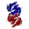
| Structure viewer | Molecule: MolmilJmol/JSmol |
|---|
- Downloads & links
-Download
| PDBx/mmCIF format | 4e08.cif.gz | 151.3 KB | Display | PDBx/mmCIF format |
|---|---|---|---|---|
| PDB format | pdb4e08.ent.gz | 120.1 KB | Display | PDB format |
| PDBx/mmJSON format | 4e08.json.gz | Tree view | PDBx/mmJSON format | |
| Others |  Other downloads Other downloads |
-Validation report
| Arichive directory | https://data.pdbj.org/pub/pdb/validation_reports/e0/4e08ftp://data.pdbj.org/pub/pdb/validation_reports/e0/4e08 | HTTPS FTP |
|---|
-Related structure data
| Related structure data | 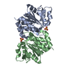 2or3S S: Starting model for refinement |
|---|---|
| Similar structure data |


-Links
 PDBj
PDBj

- Assembly
| Deposited unit | 
| ||||||||||||
|---|---|---|---|---|---|---|---|---|---|---|---|---|---|
| 1 |
| ||||||||||||
| Unit cell |
| ||||||||||||
| Components on special symmetry positions |
| ||||||||||||
| Noncrystallographic symmetry (NCS) | NCS oper:
|
-Components
| #1: Protein | Mass: 19551.367 Da / Num. of mol.: 2 Source method: isolated from a genetically manipulated source Source: (gene. exp.)  #2: Chemical | ChemComp-SO4 / |   Mass: 96.063 Da / Num. of mol.: 1 / Source method: obtained synthetically / Formula: SO4 Mass: 96.063 Da / Num. of mol.: 1 / Source method: obtained synthetically / Formula: SO4#3: Water | ChemComp-HOH / |  Mass: 18.015 Da / Num. of mol.: 278 / Source method: isolated from a natural source / Formula: H2O Mass: 18.015 Da / Num. of mol.: 278 / Source method: isolated from a natural source / Formula: H2O |
|---|
-Experimental details
-Experiment
| Experiment | Method: X-RAY DIFFRACTION / Number of used crystals: 1 |
|---|
- Sample preparation
| Crystal | Density Matthews: 2.31 Å3/Da / Density % sol: 46.82 % |
|---|---|
| Crystal grow | Temperature: 298 K / Method: vapor diffusion, sitting drop / pH: 4.6 Details: 25% polyethylene glycol 4000, 0.2 M ammonium sulfate, 0.1 M sodium acetate pH=4.6, VAPOR DIFFUSION, SITTING DROP, temperature 298K |
-Data collection
| Diffraction | Mean temperature: 110 K |
|---|---|
| Diffraction source | Source: SYNCHROTRON / Site: APS  / Beamline: 23-ID-D / Wavelength: 0.98 Å / Beamline: 23-ID-D / Wavelength: 0.98 Å |
| Detector | Type: MARMOSAIC 300 mm CCD / Detector: CCD / Date: Jul 6, 2010 / Details: K-B PAIR OF BIOMORPH MIRRORS |
| Radiation | Monochromator: DOUBLE CRYSTAL / Protocol: SINGLE WAVELENGTH / Monochromatic (M) / Laue (L): M / Scattering type: x-ray |
| Radiation wavelength | Wavelength: 0.98 Å / Relative weight: 1 |
| Reflection | Resolution: 2→76 Å / Num. all: 25116 / Num. obs: 25116 / % possible obs: 97.3 % / Observed criterion σ(F): 0 / Observed criterion σ(I): 0 / Redundancy: 8.5 % / Biso Wilson estimate: 26.9 Å2 / Rsym value: 0.098 / Net I/σ(I): 21.1 |
| Reflection shell | Resolution: 2→2.07 Å / Redundancy: 5.1 % / Mean I/σ(I) obs: 1.8 / Num. unique all: 2112 / Rsym value: 0.587 / % possible all: 83.9 |
- Processing
| Software |
| |||||||||||||||||||||||||||||||||||||||||||||||||||||||||||||||||||||||||||
|---|---|---|---|---|---|---|---|---|---|---|---|---|---|---|---|---|---|---|---|---|---|---|---|---|---|---|---|---|---|---|---|---|---|---|---|---|---|---|---|---|---|---|---|---|---|---|---|---|---|---|---|---|---|---|---|---|---|---|---|---|---|---|---|---|---|---|---|---|---|---|---|---|---|---|---|---|
| Refinement | Method to determine structure: MOLECULAR REPLACEMENT Starting model: 2OR3 Resolution: 2→75.72 Å / Cor.coef. Fo:Fc: 0.955 / Cor.coef. Fo:Fc free: 0.926 / SU B: 9.18 / SU ML: 0.132 / Cross valid method: THROUGHOUT / σ(F): 0 / ESU R: 0.197 / ESU R Free: 0.178 / Stereochemistry target values: MAXIMUM LIKELIHOOD / Details: U VALUES: WITH TLS ADDED
| |||||||||||||||||||||||||||||||||||||||||||||||||||||||||||||||||||||||||||
| Solvent computation | Ion probe radii: 0.8 Å / Shrinkage radii: 0.8 Å / VDW probe radii: 1.2 Å / Solvent model: MASK | |||||||||||||||||||||||||||||||||||||||||||||||||||||||||||||||||||||||||||
| Displacement parameters | Biso mean: 31.734 Å2
| |||||||||||||||||||||||||||||||||||||||||||||||||||||||||||||||||||||||||||
| Refinement step | Cycle: LAST / Resolution: 2→75.72 Å
| |||||||||||||||||||||||||||||||||||||||||||||||||||||||||||||||||||||||||||
| Refine LS restraints |
| |||||||||||||||||||||||||||||||||||||||||||||||||||||||||||||||||||||||||||
| LS refinement shell | Resolution: 1.999→2.051 Å / Total num. of bins used: 20
| |||||||||||||||||||||||||||||||||||||||||||||||||||||||||||||||||||||||||||
| Refinement TLS params. | Method: refined / Refine-ID: X-RAY DIFFRACTION
| |||||||||||||||||||||||||||||||||||||||||||||||||||||||||||||||||||||||||||
| Refinement TLS group |
|
