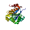
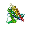
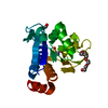
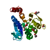
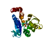
Mass: 20208.576 Da / Num. of mol.: 1 Source method: isolated from a genetically manipulated source Source: (gene. exp.) Homo sapiens (human) / Gene: PARK7 / Plasmid: SpeedET / Production host: Escherichia Coli (E. coli) / Strain (production host): PB1 References: UniProt: Q99497, Hydrolases; Acting on ester bonds; Thioester hydrolases, Hydrolases; Acting on carbon-nitrogen bonds, other than peptide bonds; In linear amides
Mass: 18.015 Da / Num. of mol.: 198 / Source method: isolated from a natural source / Formula: H2O
Has protein modification
Y
Sequence details
THE CONSTRUCT WAS EXPRESSED WITH A PURIFICATION TAG MGSDKIHHHHHHENLYFQG. THE TAG WAS REMOVED WITH ...THE CONSTRUCT WAS EXPRESSED WITH A PURIFICATION TAG MGSDKIHHHHHHENLYFQG. THE TAG WAS REMOVED WITH TEV PROTEASE LEAVING ONLY A GLYCINE (0) FOLLOWED BY THE TARGET SEQUENCE.
-
Experimental details
-
Experiment
Experiment
Method: X-RAY DIFFRACTION / Number of used crystals: 1
-
Sample preparation
Crystal
Density Matthews: 3.01 Å3/Da / Density % sol: 59.13 %
Crystal grow
Temperature: 277 K / Method: vapor diffusion, sitting drop / pH: 8 / Details: 5.00% polyethylene glycol 6000, 0.1M TRIS pH 8.0
Method to determine structure: SAD / Resolution: 1.23→29.796 Å / Cor.coef. Fo:Fc: 0.982 / Cor.coef. Fo:Fc free: 0.976 / Occupancy max: 1 / Occupancy min: 0.2 / SU B: 0.931 / SU ML: 0.017 / Cross valid method: THROUGHOUT / σ(F): 0 / ESU R: 0.027 / ESU R Free: 0.027 Stereochemistry target values: MAXIMUM LIKELIHOOD WITH PHASES Details: 1. A MET-INHIBITION PROTOCOL WAS USED FOR SELENOMETHIONINE INCORPORATION DURING PROTEIN EXPRESSION. THE OCCUPANCY OF THE SE ATOMS IN THE MSE RESIDUES WAS REDUCED TO 0.75 FOR THE REDUCED ...Details: 1. A MET-INHIBITION PROTOCOL WAS USED FOR SELENOMETHIONINE INCORPORATION DURING PROTEIN EXPRESSION. THE OCCUPANCY OF THE SE ATOMS IN THE MSE RESIDUES WAS REDUCED TO 0.75 FOR THE REDUCED SCATTERING POWER DUE TO PARTIAL S-MET INCORPORATION. 2. THE SAD PHASES WERE USED AS RESTRAINTS DURING REFINEMENT. 3. HYDROGENS HAVE BEEN ADDED IN THE RIDING POSITIONS. 4. EDO MODELED WAS PRESENT IN CRYO CONDITION.
Rfactor
Num. reflection
% reflection
Selection details
Rfree
0.1339
3374
4.8 %
RANDOM
Rwork
0.1164
66326
-
-
obs
0.1172
69700
98.23 %
-
Solvent computation
Ion probe radii: 0.8 Å / Shrinkage radii: 0.8 Å / VDW probe radii: 1.2 Å / Solvent model: MASK
In the structure databanks used in Yorodumi, some data are registered as the other names, "COVID-19 virus" and "2019-nCoV". Here are the details of the virus and the list of structure data.
Jan 31, 2019. EMDB accession codes are about to change! (news from PDBe EMDB page)
EMDB accession codes are about to change! (news from PDBe EMDB page)
The allocation of 4 digits for EMDB accession codes will soon come to an end. Whilst these codes will remain in use, new EMDB accession codes will include an additional digit and will expand incrementally as the available range of codes is exhausted. The current 4-digit format prefixed with “EMD-” (i.e. EMD-XXXX) will advance to a 5-digit format (i.e. EMD-XXXXX), and so on. It is currently estimated that the 4-digit codes will be depleted around Spring 2019, at which point the 5-digit format will come into force.
The EM Navigator/Yorodumi systems omit the EMD- prefix.
Related info.:Q: What is EMD? / ID/Accession-code notation in Yorodumi/EM Navigator
Yorodumi is a browser for structure data from EMDB, PDB, SASBDB, etc.
This page is also the successor to EM Navigator detail page, and also detail information page/front-end page for Omokage search.
The word "yorodu" (or yorozu) is an old Japanese word meaning "ten thousand". "mi" (miru) is to see.
Related info.:EMDB / PDB / SASBDB / Comparison of 3 databanks / Yorodumi Search / Aug 31, 2016. New EM Navigator & Yorodumi / Yorodumi Papers / Jmol/JSmol / Function and homology information / Changes in new EM Navigator and Yorodumi
 Movie
Movie Controller

 Yorodumi
 Open data
 Basic information
 Components
 Keywords
 Function and homology information
 Homo sapiens (human)
 X-RAY DIFFRACTION /
  Authors
 Citation
 Structure visualization
 Downloads & links
 Other downloads
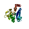


 PDBj
PDBj


 Assembly


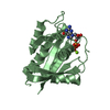
 Mass: 62.068 Da / Num. of mol.: 10 / Source method: obtained synthetically / Formula: C2H6O2
 Mass: 18.015 Da / Num. of mol.: 198 / Source method: isolated from a natural source / Formula: H2O
 Sample preparation
 / Beamline: BL14-1
 Processing