 Movie
Movie Controller


+ Open data

- Basic information
| Entry | Database: PDB / ID: 4r1b | ||||||
|---|---|---|---|---|---|---|---|
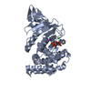
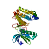
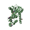
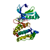
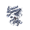
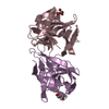
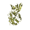
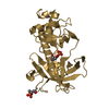
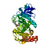
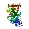
| Title | Crystal Structure of 3D7 strain Plasmodium falciparum AMA1 | ||||||
 Components Components | Apical membrane antigen 1, AMA1 | ||||||
 Keywords Keywords | CELL INVASION / PAN fold / erythrocyte invasion by merozoites / RON2 / Moving junction complex | ||||||
| Function / homology |  Function and homology information Function and homology informationapical complex / microneme / host cell surface binding / symbiont entry into host / membrane Similarity search - Function | ||||||
| Biological species |  | ||||||
| Method |  X-RAY DIFFRACTION / SYNCHROTRON / MOLECULAR REPLACEMENT / Resolution: 1.6 Å X-RAY DIFFRACTION / SYNCHROTRON / MOLECULAR REPLACEMENT / Resolution: 1.6 Å | ||||||
 Authors Authors | Lim, S.S. / Norton, R.S. / McGowan, S. | ||||||
 Citation Citation | Journal: Biochemistry / Year: 2014 Title: Structure and Dynamics of Apical Membrane Antigen 1 from Plasmodium falciparum FVO. Authors: Lim, S.S. / Yang, W. / Krishnarjuna, B. / Kannan Sivaraman, K. / Chandrashekaran, I.R. / Kass, I. / MacRaild, C.A. / Devine, S.M. / Debono, C.O. / Anders, R.F. / Scanlon, M.J. / Scammells, P. ...Authors: Lim, S.S. / Yang, W. / Krishnarjuna, B. / Kannan Sivaraman, K. / Chandrashekaran, I.R. / Kass, I. / MacRaild, C.A. / Devine, S.M. / Debono, C.O. / Anders, R.F. / Scanlon, M.J. / Scammells, P.J. / Norton, R.S. / McGowan, S. | ||||||
| History |
|
- Structure visualization
| Structure viewer | Molecule: MolmilJmol/JSmol |
|---|
- Downloads & links
-Download
| PDBx/mmCIF format | 4r1b.cif.gz | 138.4 KB | Display | PDBx/mmCIF format |
|---|---|---|---|---|
| PDB format | pdb4r1b.ent.gz | 107.3 KB | Display | PDB format |
| PDBx/mmJSON format | 4r1b.json.gz | Tree view | PDBx/mmJSON format | |
| Others |  Other downloads Other downloads |
-Validation report
| Arichive directory | https://data.pdbj.org/pub/pdb/validation_reports/r1/4r1bftp://data.pdbj.org/pub/pdb/validation_reports/r1/4r1b | HTTPS FTP |
|---|
-Related structure data
| Related structure data | 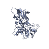 4r19C  4r1aC 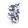 4r1cC 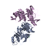 1z40S C: citing same article ( S: Starting model for refinement |
|---|---|
| Similar structure data |
-Links
 PDBj
PDBj- Assembly
| Deposited unit | 
| ||||||||
|---|---|---|---|---|---|---|---|---|---|
| 1 |
| ||||||||
| Unit cell |
| ||||||||
| Components on special symmetry positions |
|
-Components
| #1: Protein | Mass: 38360.047 Da / Num. of mol.: 1 / Fragment: UNP residues 104-438 Source method: isolated from a genetically manipulated source Source: (gene. exp.)  |
|---|---|
| #2: Water | ChemComp-HOH /  Mass: 18.015 Da / Num. of mol.: 296 / Source method: isolated from a natural source / Formula: H2O Mass: 18.015 Da / Num. of mol.: 296 / Source method: isolated from a natural source / Formula: H2O |
| Has protein modification | Y |
-Experimental details
-Experiment
| Experiment | Method: X-RAY DIFFRACTION / Number of used crystals: 1 |
|---|
- Sample preparation
| Crystal | Density Matthews: 2 Å3/Da / Density % sol: 38.61 % |
|---|---|
| Crystal grow | Temperature: 298 K / Method: vapor diffusion, hanging drop / pH: 6 Details: 12-15% (v/v) polyethylene glycol 3350, 0.02 M MES and 10 mM manganese chloride. Crystals were dehydrated overnight in reservoir solution with increased (35%) (v/v) polyethylene glycol 3350 ...Details: 12-15% (v/v) polyethylene glycol 3350, 0.02 M MES and 10 mM manganese chloride. Crystals were dehydrated overnight in reservoir solution with increased (35%) (v/v) polyethylene glycol 3350 before cryo-stabilisation in 38% (v/v) polyethylene glycol 3350, 0.088 M MES (pH 6.0) and 44 mM manganese chloride for 6-8 h prior to data collection. MilliQ water was added to the stabilisation solution, VAPOR DIFFUSION, HANGING DROP, temperature 298K |
-Data collection
| Diffraction | Mean temperature: 100 K |
|---|---|
| Diffraction source | Source: SYNCHROTRON / Site: Australian Synchrotron  / Beamline: MX2 / Wavelength: 0.9537 Å / Beamline: MX2 / Wavelength: 0.9537 Å |
| Detector | Type: ADSC QUANTUM 315r / Detector: CCD / Date: Nov 30, 2013 |
| Radiation | Monochromator: DOUBLE CRYSTAL SILICON 111 / Protocol: SINGLE WAVELENGTH / Monochromatic (M) / Laue (L): M / Scattering type: x-ray |
| Radiation wavelength | Wavelength: 0.9537 Å / Relative weight: 1 |
| Reflection | Resolution: 1.6→41.07 Å / Num. obs: 40241 / % possible obs: 100 % / Observed criterion σ(I): 8 |
- Processing
| Software |
| |||||||||||||||||||||||||||||||||||||||||||||||||||||||||||||||||||||||||||||||||||||||||||||||||||||||||
|---|---|---|---|---|---|---|---|---|---|---|---|---|---|---|---|---|---|---|---|---|---|---|---|---|---|---|---|---|---|---|---|---|---|---|---|---|---|---|---|---|---|---|---|---|---|---|---|---|---|---|---|---|---|---|---|---|---|---|---|---|---|---|---|---|---|---|---|---|---|---|---|---|---|---|---|---|---|---|---|---|---|---|---|---|---|---|---|---|---|---|---|---|---|---|---|---|---|---|---|---|---|---|---|---|---|---|
| Refinement | Method to determine structure: MOLECULAR REPLACEMENT Starting model: PDB ID: 1Z40 Resolution: 1.6→37.454 Å / SU ML: 0.17 / σ(F): 1.34 / Phase error: 21.22 / Stereochemistry target values: ML
| |||||||||||||||||||||||||||||||||||||||||||||||||||||||||||||||||||||||||||||||||||||||||||||||||||||||||
| Solvent computation | Shrinkage radii: 0.9 Å / VDW probe radii: 1.11 Å / Solvent model: FLAT BULK SOLVENT MODEL | |||||||||||||||||||||||||||||||||||||||||||||||||||||||||||||||||||||||||||||||||||||||||||||||||||||||||
| Refinement step | Cycle: LAST / Resolution: 1.6→37.454 Å
| |||||||||||||||||||||||||||||||||||||||||||||||||||||||||||||||||||||||||||||||||||||||||||||||||||||||||
| Refine LS restraints |
| |||||||||||||||||||||||||||||||||||||||||||||||||||||||||||||||||||||||||||||||||||||||||||||||||||||||||
| LS refinement shell |
| |||||||||||||||||||||||||||||||||||||||||||||||||||||||||||||||||||||||||||||||||||||||||||||||||||||||||
| Refinement TLS params. | Method: refined / Origin x: -26.3118 Å / Origin y: 11.5211 Å / Origin z: 104.7439 Å
| |||||||||||||||||||||||||||||||||||||||||||||||||||||||||||||||||||||||||||||||||||||||||||||||||||||||||
| Refinement TLS group | Selection details: all |
