 Movie
Movie Controller

[English] 日本語
 Yorodumi
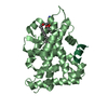
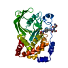
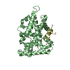
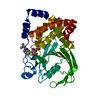
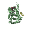
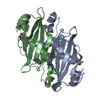
- PDB-4qk4: Crystal structure of human nuclear receptor sf-1 (nr5a1) bound to... -
+ Open data

- Basic information
| Entry | Database: PDB / ID: 4qk4 | ||||||
|---|---|---|---|---|---|---|---|
| Title | Crystal structure of human nuclear receptor sf-1 (nr5a1) bound to pip2 at 2.8 a resolution | ||||||
 Components Components |
| ||||||
 Keywords Keywords | TRANSCRIPTION FACTOR/HORMONE / NUCLEAR HORMONE RECEPTOR / NR5A1 / SF-1 LIGAND BINDING DOMAIN / REGULATORY LIGANDS / TRANSCRIPTION / TRANSCRIPTION REGULATION / STRUCTURAL GENOMICS / JOINT CENTER FOR STRUCTURAL GENOMICS / JCSG / PROTEIN STRUCTURE INITIATIVE / PSI-BIOLOGY / PARTNERSHIP FOR STEM CELL BIOLOGY / PIP3 / PIP2 / NUCLEUS / NUCLEAR PHOSPHATIDYLINOSITOL PHOSPHATES / TRANSCRIPTION FACTOR-HORMONE complex / STEMCELL | ||||||
| Function / homology |  Function and homology information Function and homology informationprimary sex determination / response to gonadotropin-releasing hormone / Sertoli cell differentiation / negative regulation of female gonad development / luteinization / regulation of steroid biosynthetic process / sex determination / positive regulation of male gonad development / Regulation of MITF-M dependent genes involved in metabolism / Transcriptional regulation of testis differentiation ...primary sex determination / response to gonadotropin-releasing hormone / Sertoli cell differentiation / negative regulation of female gonad development / luteinization / regulation of steroid biosynthetic process / sex determination / positive regulation of male gonad development / Regulation of MITF-M dependent genes involved in metabolism / Transcriptional regulation of testis differentiation / tissue development / Transcriptional regulation of pluripotent stem cells / Leydig cell differentiation / positive regulation of fatty acid oxidation / male sex determination / cellular respiration / maintenance of protein location in nucleus / hormone metabolic process / Activation of PPARGC1A (PGC-1alpha) by phosphorylation / adrenal gland development / response to starvation / female gonad development / response to muscle activity / temperature homeostasis / lncRNA binding / response to dietary excess / fatty acid oxidation / intracellular glucose homeostasis / calcineurin-mediated signaling / adipose tissue development / FOXO-mediated transcription of oxidative stress, metabolic and neuronal genes / Transcriptional regulation of brown and beige adipocyte differentiation by EBF2 / energy homeostasis / digestion / : / hormone-mediated signaling pathway / brown fat cell differentiation / positive regulation of gluconeogenesis / SUMOylation of transcription cofactors / RNA splicing / nuclear receptor binding / transcription coregulator binding / gluconeogenesis / respiratory electron transport chain / RNA polymerase II transcription regulatory region sequence-specific DNA binding / transcription coregulator activity / mitochondrion organization / SUMOylation of intracellular receptors / circadian regulation of gene expression / transcription initiation at RNA polymerase II promoter / negative regulation of smooth muscle cell proliferation / Heme signaling / PPARA activates gene expression / Transcriptional activation of mitochondrial biogenesis / regulation of circadian rhythm / chromatin DNA binding / PML body / phospholipid binding / Nuclear Receptor transcription pathway / Transcriptional regulation of white adipocyte differentiation / RNA polymerase II transcription regulator complex / male gonad development / nuclear receptor activity / mRNA processing / sequence-specific double-stranded DNA binding / Regulation of RUNX2 expression and activity / positive regulation of cold-induced thermogenesis / MLL4 and MLL3 complexes regulate expression of PPARG target genes in adipogenesis and hepatic steatosis / cellular response to oxidative stress / protein-containing complex assembly / neuron apoptotic process / sequence-specific DNA binding / DNA-binding transcription factor binding / transcription by RNA polymerase II / negative regulation of neuron apoptotic process / RNA polymerase II-specific DNA-binding transcription factor binding / DNA-binding transcription factor activity, RNA polymerase II-specific / transcription coactivator activity / protein stabilization / RNA polymerase II cis-regulatory region sequence-specific DNA binding / ubiquitin protein ligase binding / chromatin binding / positive regulation of gene expression / regulation of transcription by RNA polymerase II / regulation of DNA-templated transcription / positive regulation of DNA-templated transcription / chromatin / enzyme binding / positive regulation of transcription by RNA polymerase II / DNA binding / RNA binding / zinc ion binding / nucleoplasm / nucleus / cytosol Similarity search - Function | ||||||
| Biological species |  Homo sapiens (human) Homo sapiens (human) | ||||||
| Method |  X-RAY DIFFRACTION / MOLECULAR REPLACEMENT / molecular replacement / Resolution: 2.81 Å X-RAY DIFFRACTION / MOLECULAR REPLACEMENT / molecular replacement / Resolution: 2.81 Å | ||||||
 Authors Authors | Joint Center for Structural Genomics (JCSG) / Partnership for Stem Cell Biology (STEMCELL) | ||||||
 Citation Citation | Journal: Proc.Natl.Acad.Sci.USA / Year: 2014 Title: The signaling phospholipid PIP3 creates a new interaction surface on the nuclear receptor SF-1. Authors: Blind, R.D. / Sablin, E.P. / Kuchenbecker, K.M. / Chiu, H.J. / Deacon, A.M. / Das, D. / Fletterick, R.J. / Ingraham, H.A. | ||||||
| History |
|
- Structure visualization
| Structure viewer | Molecule: MolmilJmol/JSmol |
|---|
- Downloads & links
-Download
| PDBx/mmCIF format | 4qk4.cif.gz | 126 KB | Display | PDBx/mmCIF format |
|---|---|---|---|---|
| PDB format | pdb4qk4.ent.gz | 98 KB | Display | PDB format |
| PDBx/mmJSON format | 4qk4.json.gz | Tree view | PDBx/mmJSON format | |
| Others |  Other downloads Other downloads |
-Validation report
| Arichive directory | https://data.pdbj.org/pub/pdb/validation_reports/qk/4qk4ftp://data.pdbj.org/pub/pdb/validation_reports/qk/4qk4 | HTTPS FTP |
|---|
-Related structure data
| Related structure data |  4qjrC  1yowS S: Starting model for refinement C: citing same article ( |
|---|---|
| Similar structure data | |
| Other databases |


-Links
 PDBj
PDBj


- Assembly
| Deposited unit | 
| ||||||||
|---|---|---|---|---|---|---|---|---|---|
| 1 |
| ||||||||
| Unit cell |
|
-Components
| #1: Protein | Mass: 27903.406 Da / Num. of mol.: 1 / Fragment: UNP residues 218-461 / Mutation: C247S, C412S Source method: isolated from a genetically manipulated source Source: (gene. exp.) Homo sapiens (human) / Gene: AD4BP, FTZF1, NR5A1, RC2003B, SF1 / Plasmid: pBH4 / Production host:  | ||||
|---|---|---|---|---|---|
| #2: Protein/peptide | Mass: 1523.854 Da / Num. of mol.: 1 / Fragment: UNP residues 139-152 / Source method: obtained synthetically Details: PGC-1ALPHA (PEROXISOME PROLIFERATOR-ACTIVATED RECEPTOR GAMMA CO-ACTIVATOR-1ALPHA) PEPTIDE CONTAINING RESIDUES 139-EEPSLLKKLLLAPA-152 Source: (synth.) Homo sapiens (human) / References: UniProt: Q9UBK2 | ||||
| #3: Chemical | ChemComp-PIK / (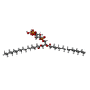  Mass: 970.992 Da / Num. of mol.: 1 / Source method: obtained synthetically / Formula: C41H81O19P3 Mass: 970.992 Da / Num. of mol.: 1 / Source method: obtained synthetically / Formula: C41H81O19P3Details: SMILES string: CCCCCCCCCCCCCCCC(=O)OCC(CO[P](O)(=O)OC1C(O)C(O)C (O[P](O)(O)=O)C(O[P](O)(O)=O)C1O)OC(=O)CCCCCCCCCCCCCCC | ||||
| #4: Chemical | ChemComp-EDO /   Mass: 62.068 Da / Num. of mol.: 5 / Source method: obtained synthetically / Formula: C2H6O2 Mass: 62.068 Da / Num. of mol.: 5 / Source method: obtained synthetically / Formula: C2H6O2#5: Water | ChemComp-HOH / |  Mass: 18.015 Da / Num. of mol.: 55 / Source method: isolated from a natural source / Formula: H2O Mass: 18.015 Da / Num. of mol.: 55 / Source method: isolated from a natural source / Formula: H2OSequence details | SF-1 (UNIPROT Q13285, NR5A1_HUMAN, STF1_HUMAN) LIGAND BINDING DOMAIN (LBD) WAS EXPRESSED WITH AN N- ...SF-1 (UNIPROT Q13285, NR5A1_HUMAN, STF1_HUMAN) LIGAND BINDING DOMAIN (LBD) WAS EXPRESSED WITH AN N-TERMINAL PURIFICATI | |
-Experimental details
-Experiment
| Experiment | Method: X-RAY DIFFRACTION / Number of used crystals: 1 |
|---|
- Sample preparation
| Crystal | Density Matthews: 3.31 Å3/Da / Density % sol: 62.87 % |
|---|---|
| Crystal grow | Temperature: 277 K / Method: vapor diffusion, sitting drop Details: SF-1, 6% PEG 8000, 0.2M MGOAC, 20% ETHYLENE GLYCOL, 0.067MM PIP2, 0.80MM 14-MER EEPSLLKKLLLAPA, NANODROP, VAPOR DIFFUSION, SITTING DROP, TEMPERATURE 277K |
-Data collection
| Diffraction | Mean temperature: 100 K | |||||||||||||||||||||||||||||||||||||||||||||||||||||||||||||||||||||||||||||
|---|---|---|---|---|---|---|---|---|---|---|---|---|---|---|---|---|---|---|---|---|---|---|---|---|---|---|---|---|---|---|---|---|---|---|---|---|---|---|---|---|---|---|---|---|---|---|---|---|---|---|---|---|---|---|---|---|---|---|---|---|---|---|---|---|---|---|---|---|---|---|---|---|---|---|---|---|---|---|
| Diffraction source | Source: ROTATING ANODE / Type: RIGAKU MICROMAX-002+ / Wavelength: 1.5418 | |||||||||||||||||||||||||||||||||||||||||||||||||||||||||||||||||||||||||||||
| Detector | Type: MARMOSAIC 325 mm CCD / Detector: CCD / Date: Sep 16, 2013 / Details: MIRROR | |||||||||||||||||||||||||||||||||||||||||||||||||||||||||||||||||||||||||||||
| Radiation | Protocol: SINGLE WAVELENGTH / Monochromatic (M) / Laue (L): M / Scattering type: x-ray | |||||||||||||||||||||||||||||||||||||||||||||||||||||||||||||||||||||||||||||
| Radiation wavelength | Wavelength: 1.5418 Å / Relative weight: 1 | |||||||||||||||||||||||||||||||||||||||||||||||||||||||||||||||||||||||||||||
| Reflection | Resolution: 2.81→29.119 Å / Num. obs: 10167 / % possible obs: 99.4 % / Observed criterion σ(I): -3 / Biso Wilson estimate: 90.29 Å2 / Rmerge(I) obs: 0.042 / Net I/σ(I): 23.72 | |||||||||||||||||||||||||||||||||||||||||||||||||||||||||||||||||||||||||||||
| Reflection shell | Diffraction-ID: 1,2
|
-Phasing
| Phasing | Method: molecular replacement |
|---|
- Processing
| Software |
| ||||||||||||||||||||||||||||||||||||||||||||||||||||||||||||||||||||||||||||||||||||||||||||||||||||
|---|---|---|---|---|---|---|---|---|---|---|---|---|---|---|---|---|---|---|---|---|---|---|---|---|---|---|---|---|---|---|---|---|---|---|---|---|---|---|---|---|---|---|---|---|---|---|---|---|---|---|---|---|---|---|---|---|---|---|---|---|---|---|---|---|---|---|---|---|---|---|---|---|---|---|---|---|---|---|---|---|---|---|---|---|---|---|---|---|---|---|---|---|---|---|---|---|---|---|---|---|---|
| Refinement | Method to determine structure: MOLECULAR REPLACEMENT Starting model: PDB entry 1YOW Resolution: 2.81→29.119 Å / Occupancy max: 1 / Occupancy min: 0.32 / SU ML: 0.43 / σ(F): 2 / Phase error: 28.14 / Stereochemistry target values: ML Details: 1. ATOM RECORD CONTAINS SUM OF TLS AND RESIDUAL B FACTORS. ANISOU RECORD CONTAINS SUM OF TLS AND RESIDUAL U FACTORS. 2. 1,2-ETHANEDIOL (EDO) FROM THE CRYSTALLIZATION SOLUTION HAS BEEN ...Details: 1. ATOM RECORD CONTAINS SUM OF TLS AND RESIDUAL B FACTORS. ANISOU RECORD CONTAINS SUM OF TLS AND RESIDUAL U FACTORS. 2. 1,2-ETHANEDIOL (EDO) FROM THE CRYSTALLIZATION SOLUTION HAS BEEN MODELED IN THE SOLVENT STRUCTURE. 3. PIP2 WAS CO-CRYSTALLIZED WITH THE SF-1 PROTEIN AND PGC-1ALPHA PEPTIDE.
| ||||||||||||||||||||||||||||||||||||||||||||||||||||||||||||||||||||||||||||||||||||||||||||||||||||
| Solvent computation | Shrinkage radii: 0.9 Å / VDW probe radii: 1.11 Å / Solvent model: FLAT BULK SOLVENT MODEL | ||||||||||||||||||||||||||||||||||||||||||||||||||||||||||||||||||||||||||||||||||||||||||||||||||||
| Displacement parameters | Biso max: 180.41 Å2 / Biso mean: 73.8761 Å2 / Biso min: 31.06 Å2 | ||||||||||||||||||||||||||||||||||||||||||||||||||||||||||||||||||||||||||||||||||||||||||||||||||||
| Refinement step | Cycle: LAST / Resolution: 2.81→29.119 Å
| ||||||||||||||||||||||||||||||||||||||||||||||||||||||||||||||||||||||||||||||||||||||||||||||||||||
| Refine LS restraints |
| ||||||||||||||||||||||||||||||||||||||||||||||||||||||||||||||||||||||||||||||||||||||||||||||||||||
| LS refinement shell | Refine-ID: X-RAY DIFFRACTION / Total num. of bins used: 4
| ||||||||||||||||||||||||||||||||||||||||||||||||||||||||||||||||||||||||||||||||||||||||||||||||||||
| Refinement TLS params. | Method: refined / Refine-ID: X-RAY DIFFRACTION
| ||||||||||||||||||||||||||||||||||||||||||||||||||||||||||||||||||||||||||||||||||||||||||||||||||||
| Refinement TLS group |
|
