 Movie
Movie Controller


+ Open data

- Basic information
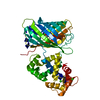
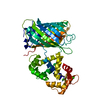
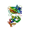
| Entry | Database: PDB / ID: 4ik5 | ||||||
|---|---|---|---|---|---|---|---|
| Title | High resolution structure of Delta-REST-GCaMP3 | ||||||
 Components Components | RCaMP, Green fluorescent protein | ||||||
 Keywords Keywords |  FLUORESCENT PROTEIN / calcium indicator / mutants / fluorescent intensity / dimerization / beta-barrel / Calmodulin FLUORESCENT PROTEIN / calcium indicator / mutants / fluorescent intensity / dimerization / beta-barrel / Calmodulin | ||||||
| Function / homology |  Function and homology informationbioluminescence / generation of precursor metabolites and energy / calcium ion binding Function and homology informationbioluminescence / generation of precursor metabolites and energy / calcium ion bindingSimilarity search - Function | ||||||
| Biological species |  Entacmaea quadricolor (sea anemone) Entacmaea quadricolor (sea anemone) Aequorea victoria (jellyfish) Aequorea victoria (jellyfish) | ||||||
| Method | X-RAY DIFFRACTION / MOLECULAR REPLACEMENT / Resolution: 2.5 Å | ||||||
 Authors Authors | Chen, Y. / Song, X. / Miao, L. / Zhu, Y. / Ji, G. | ||||||
 Citation Citation | Journal: Protein Cell / Year: 2013 Title: Structural insight into enhanced calcium indicator GCaMP3 and GCaMPJ to promote further improvement. Authors: Chen, Y. / Song, X. / Ye, S. / Miao, L. / Zhu, Y. / Zhang, R.G. / Ji, G. | ||||||
| History |
|
- Structure visualization
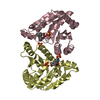
| Structure viewer | Molecule: MolmilJmol/JSmol |
|---|
- Downloads & links
-Download
| PDBx/mmCIF format | 4ik5.cif.gz | 97.6 KB | Display | PDBx/mmCIF format |
|---|---|---|---|---|
| PDB format | pdb4ik5.ent.gz | 77.2 KB | Display | PDB format |
| PDBx/mmJSON format | 4ik5.json.gz | Tree view | PDBx/mmJSON format | |
| Others |  Other downloads Other downloads |
-Validation report
| Arichive directory | https://data.pdbj.org/pub/pdb/validation_reports/ik/4ik5ftp://data.pdbj.org/pub/pdb/validation_reports/ik/4ik5 | HTTPS FTP |
|---|
-Related structure data


-Links
 PDBj
PDBj


- Assembly
| Deposited unit | 
| |||||||||
|---|---|---|---|---|---|---|---|---|---|---|
| 1 |
| |||||||||
| Unit cell |
| |||||||||
| Components on special symmetry positions |
|
-Components
| #1: Protein | Mass: 46764.324 Da / Num. of mol.: 1 Mutation: M153K, V163A, S175G, D180Y, T203V, A206K, H231L, F64L, V93I, I354T Source method: isolated from a genetically manipulated source Details: CHIMERA OF RESIDUES 25-48 OF RCaMP, LINKER LE, RESIDUES 149-238 OF GREEN FLUORESCENT PROTEIN, LINKER GGTGGSMV, RESIDUES 2-144 OF GREEN FLUORESCENT PROTEIN, RESIDUES 284-432 OF RCaMP Source: (gene. exp.) Entacmaea quadricolor (sea anemone), (gene. exp.) Aequorea victoria (jellyfish)Gene: GFP / Production host:  Escherichia coli (E. coli) / References: UniProt: K4DIE3, UniProt: P42212 Escherichia coli (E. coli) / References: UniProt: K4DIE3, UniProt: P42212 | ||||
|---|---|---|---|---|---|
| #2: Chemical | ChemComp-CA /   Mass: 40.078 Da / Num. of mol.: 4 / Source method: obtained synthetically / Formula: Ca Mass: 40.078 Da / Num. of mol.: 4 / Source method: obtained synthetically / Formula: Ca#3: Water | ChemComp-HOH / | Water Mass: 18.015 Da / Num. of mol.: 306 / Source method: isolated from a natural source / Formula: H2O Mass: 18.015 Da / Num. of mol.: 306 / Source method: isolated from a natural source / Formula: H2OSequence details | (1) THIS CHIMERA COMPRISES OF RESIDUES 25-48 OF RCaMP, LINKER LE, RESIDUES 149-238 OF GREEN ...(1) THIS CHIMERA COMPRISES OF RESIDUES 25-48 OF RCaMP, LINKER LE, RESIDUES 149-238 OF GREEN FLUORESCEN | |
-Experimental details
-Experiment
| Experiment | Method: X-RAY DIFFRACTION / Number of used crystals: 1 |
|---|
- Sample preparation
| Crystal | Density Matthews: 3.73 Å3/Da / Density % sol: 67.03 % |
|---|---|
| Crystal grow | Temperature: 289 K / Method: vapor diffusion, hanging drop / pH: 8.5 Details: Tris pH 8.5, NH4OAc, 23% PEG 3350, VAPOR DIFFUSION, HANGING DROP, temperature 289K |
-Data collection
| Diffraction | Mean temperature: 100 K |
|---|---|
| Diffraction source | Source: ROTATING ANODE / Type: RIGAKU MICROMAX-007 / Wavelength: 1.5418 Å |
| Detector | Type: RIGAKU RAXIS IV / Detector: IMAGE PLATE / Date: May 3, 2012 |
| Radiation | Monochromator: Be filter / Protocol: SINGLE WAVELENGTH / Monochromatic (M) / Laue (L): M / Scattering type: x-ray |
| Radiation wavelength | Wavelength: 1.5418 Å / Relative weight: 1 |
| Reflection | Resolution: 2.4→46.95 Å / Num. all: 28413 / Num. obs: 27021 / % possible obs: 95.1 % / Observed criterion σ(F): 1 / Observed criterion σ(I): 1 |
| Reflection shell | Resolution: 2.4→2.47 Å / % possible all: 47.1 |
- Processing
| Software |
| |||||||||||||||||||||||||
|---|---|---|---|---|---|---|---|---|---|---|---|---|---|---|---|---|---|---|---|---|---|---|---|---|---|---|
| Refinement | Method to determine structure: MOLECULAR REPLACEMENT / Resolution: 2.5→46.95 Å / Cor.coef. Fo:Fc: 0.951 / Cor.coef. Fo:Fc free: 0.924 / SU B: 6.655 / SU ML: 0.15 / Cross valid method: THROUGHOUT / ESU R: 0.266 / ESU R Free: 0.222 / Stereochemistry target values: MAXIMUM LIKELIHOOD / Details: HYDROGENS HAVE BEEN ADDED IN THE RIDING POSITIONS
| |||||||||||||||||||||||||
| Solvent computation | Ion probe radii: 0.8 Å / Shrinkage radii: 0.8 Å / VDW probe radii: 1.4 Å / Solvent model: MASK | |||||||||||||||||||||||||
| Displacement parameters | Biso mean: 42.677 Å2
| |||||||||||||||||||||||||
| Refinement step | Cycle: LAST / Resolution: 2.5→46.95 Å
| |||||||||||||||||||||||||
| Refine LS restraints |
| |||||||||||||||||||||||||
| LS refinement shell | Resolution: 2.5→2.565 Å / Total num. of bins used: 20
|
