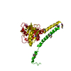
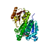
Crystal structure of Trichinella spiralis UCH37 catalytic domain bound to Ubiquitin vinyl methyl ester
Components
Ubiquitin
Ubiquitin carboxyl-hydrolase
Keywords
Hydrolase/Signaling Protein / helix-beta-helix sandwich / protein-protein complex / deubiquitination / ubiquitin c-terminal hydrolase / cytosol / Hydrolase-Signaling Protein complex
Function / homology
Function and homology information
Maturation of protein E / Maturation of protein E / ER Quality Control Compartment (ERQC) / Myoclonic epilepsy of Lafora / FLT3 signaling by CBL mutants / Constitutive Signaling by NOTCH1 HD Domain Mutants / IRAK2 mediated activation of TAK1 complex / Prevention of phagosomal-lysosomal fusion / Alpha-protein kinase 1 signaling pathway / Glycogen synthesis ...Maturation of protein E / Maturation of protein E / ER Quality Control Compartment (ERQC) / Myoclonic epilepsy of Lafora / FLT3 signaling by CBL mutants / Constitutive Signaling by NOTCH1 HD Domain Mutants / IRAK2 mediated activation of TAK1 complex / Prevention of phagosomal-lysosomal fusion / Alpha-protein kinase 1 signaling pathway / Glycogen synthesis / IRAK1 recruits IKK complex / IRAK1 recruits IKK complex upon TLR7/8 or 9 stimulation / Endosomal Sorting Complex Required For Transport (ESCRT) / Membrane binding and targetting of GAG proteins / Negative regulation of FLT3 / Regulation of TBK1, IKKε (IKBKE)-mediated activation of IRF3, IRF7 / PTK6 Regulates RTKs and Their Effectors AKT1 and DOK1 / Regulation of TBK1, IKKε-mediated activation of IRF3, IRF7 upon TLR3 ligation / IRAK2 mediated activation of TAK1 complex upon TLR7/8 or 9 stimulation / NOTCH2 Activation and Transmission of Signal to the Nucleus / TICAM1,TRAF6-dependent induction of TAK1 complex / TICAM1-dependent activation of IRF3/IRF7 / APC/C:Cdc20 mediated degradation of Cyclin B / Regulation of FZD by ubiquitination / Downregulation of ERBB4 signaling / APC-Cdc20 mediated degradation of Nek2A / p75NTR recruits signalling complexes / InlA-mediated entry of Listeria monocytogenes into host cells / TRAF6 mediated IRF7 activation in TLR7/8 or 9 signaling / Regulation of pyruvate metabolism / NF-kB is activated and signals survival / TRAF6-mediated induction of TAK1 complex within TLR4 complex / Downregulation of ERBB2:ERBB3 signaling / Pexophagy / Regulation of innate immune responses to cytosolic DNA / NRIF signals cell death from the nucleus / Regulation of PTEN localization / Activated NOTCH1 Transmits Signal to the Nucleus / VLDLR internalisation and degradation / Synthesis of active ubiquitin: roles of E1 and E2 enzymes / TICAM1, RIP1-mediated IKK complex recruitment / Regulation of BACH1 activity / Translesion synthesis by REV1 / MAP3K8 (TPL2)-dependent MAPK1/3 activation / Degradation of CDH1 / Translesion synthesis by POLK / InlB-mediated entry of Listeria monocytogenes into host cell / JNK (c-Jun kinases) phosphorylation and activation mediated by activated human TAK1 / Activation of IRF3, IRF7 mediated by TBK1, IKKε (IKBKE) / Josephin domain DUBs / Downregulation of TGF-beta receptor signaling / Translesion synthesis by POLI / Gap-filling DNA repair synthesis and ligation in GG-NER / IKK complex recruitment mediated by RIP1 / Degradation of CRY and PER proteins / Regulation of activated PAK-2p34 by proteasome mediated degradation / PINK1-PRKN Mediated Mitophagy / TGF-beta receptor signaling in EMT (epithelial to mesenchymal transition) / TNFR1-induced NF-kappa-B signaling pathway / Autodegradation of Cdh1 by Cdh1:APC/C / TCF dependent signaling in response to WNT / Regulation of NF-kappa B signaling / APC/C:Cdc20 mediated degradation of Securin / N-glycan trimming in the ER and Calnexin/Calreticulin cycle / activated TAK1 mediates p38 MAPK activation / Asymmetric localization of PCP proteins / Ubiquitin-dependent degradation of Cyclin D / SCF-beta-TrCP mediated degradation of Emi1 / NIK-->noncanonical NF-kB signaling / Regulation of signaling by CBL / TNFR2 non-canonical NF-kB pathway / AUF1 (hnRNP D0) binds and destabilizes mRNA / Negative regulators of DDX58/IFIH1 signaling / NOTCH3 Activation and Transmission of Signal to the Nucleus / Assembly of the pre-replicative complex / Negative regulation of FGFR3 signaling / Fanconi Anemia Pathway / Peroxisomal protein import / Vpu mediated degradation of CD4 / Deactivation of the beta-catenin transactivating complex / Stabilization of p53 / Degradation of DVL / Cdc20:Phospho-APC/C mediated degradation of Cyclin A / Negative regulation of FGFR2 signaling / Dectin-1 mediated noncanonical NF-kB signaling / Negative regulation of FGFR4 signaling / Downregulation of SMAD2/3:SMAD4 transcriptional activity / Degradation of AXIN / Negative regulation of FGFR1 signaling / Regulation of TNFR1 signaling / Hh mutants are degraded by ERAD / EGFR downregulation / Termination of translesion DNA synthesis / SMAD2/SMAD3:SMAD4 heterotrimer regulates transcription / Activation of NF-kappaB in B cells / G2/M Checkpoints / Assembly Of The HIV Virion / Hedgehog ligand biogenesis / Degradation of GLI1 by the proteasome / Defective CFTR causes cystic fibrosis Similarity search - Function
Method to determine structure: SAD / Resolution: 1.701→27.88 Å / SU ML: 0.2 / Cross valid method: THROUGHOUT / σ(F): 1.34 / Phase error: 25.4 / Stereochemistry target values: ML
Rfactor
Num. reflection
% reflection
Selection details
Rfree
0.2114
3133
5.03 %
random
Rwork
0.1744
-
-
-
obs
0.1763
62257
88.35 %
-
all
-
70466
-
-
Solvent computation
Shrinkage radii: 0.9 Å / VDW probe radii: 1.11 Å / Solvent model: FLAT BULK SOLVENT MODEL
Refinement step
Cycle: LAST / Resolution: 1.701→27.88 Å
Protein
Nucleic acid
Ligand
Solvent
Total
Num. atoms
4619
0
25
346
4990
Refine LS restraints
Refine-ID
Type
Dev ideal
Number
X-RAY DIFFRACTION
f_bond_d
0.017
4767
X-RAY DIFFRACTION
f_angle_d
1.753
6451
X-RAY DIFFRACTION
f_dihedral_angle_d
15.647
1787
X-RAY DIFFRACTION
f_chiral_restr
0.122
736
X-RAY DIFFRACTION
f_plane_restr
0.008
830
LS refinement shell
Resolution (Å)
Rfactor Rfree
Num. reflection Rfree
Rfactor Rwork
Num. reflection Rwork
Refine-ID
% reflection obs (%)
1.7006-1.7272
0.293
59
0.276
1235
X-RAY DIFFRACTION
41
1.7272-1.7555
0.3321
75
0.2456
1431
X-RAY DIFFRACTION
47
1.7555-1.7858
0.2836
66
0.2369
1666
X-RAY DIFFRACTION
54
1.7858-1.8183
0.2436
87
0.2427
1839
X-RAY DIFFRACTION
61
1.8183-1.8532
0.3021
128
0.2363
2193
X-RAY DIFFRACTION
72
1.8532-1.891
0.3034
112
0.242
2474
X-RAY DIFFRACTION
82
1.891-1.9321
0.2539
157
0.234
2771
X-RAY DIFFRACTION
92
1.9321-1.9771
0.2652
162
0.2091
2857
X-RAY DIFFRACTION
96
1.9771-2.0265
0.2355
178
0.1948
2990
X-RAY DIFFRACTION
99
2.0265-2.0813
0.242
163
0.1845
2976
X-RAY DIFFRACTION
99
2.0813-2.1425
0.2418
151
0.1861
3088
X-RAY DIFFRACTION
100
2.1425-2.2116
0.2246
177
0.1781
2992
X-RAY DIFFRACTION
100
2.2116-2.2906
0.235
148
0.1762
3035
X-RAY DIFFRACTION
100
2.2906-2.3823
0.2402
158
0.1818
3057
X-RAY DIFFRACTION
100
2.3823-2.4907
0.2417
172
0.1816
3019
X-RAY DIFFRACTION
100
2.4907-2.6219
0.2445
167
0.1903
3028
X-RAY DIFFRACTION
100
2.6219-2.786
0.2463
169
0.1934
3056
X-RAY DIFFRACTION
100
2.786-3.0009
0.2099
170
0.19
3009
X-RAY DIFFRACTION
100
3.0009-3.3025
0.2155
146
0.1763
3087
X-RAY DIFFRACTION
100
3.3025-3.7793
0.1785
150
0.1444
3070
X-RAY DIFFRACTION
100
3.7793-4.7577
0.1586
171
0.1277
3080
X-RAY DIFFRACTION
100
4.7577-27.8832
0.1667
167
0.1633
3171
X-RAY DIFFRACTION
100
Refinement TLS params.
Method: refined / Origin x: 35.4692 Å / Origin y: 51.0532 Å / Origin z: 17.3875 Å
11
12
13
21
22
23
31
32
33
T
0.2773 Å2
0.0143 Å2
0.0083 Å2
-
0.2452 Å2
0.008 Å2
-
-
0.264 Å2
L
0.7776 °2
-0.2111 °2
1.1152 °2
-
0.0699 °2
-0.4289 °2
-
-
1.7801 °2
S
0.0156 Å °
0.1582 Å °
0.0021 Å °
-0.023 Å °
-0.0525 Å °
-0.0208 Å °
-0.0073 Å °
0.233 Å °
0.0413 Å °
Refinement TLS group
Selection details: all
+
About Yorodumi
-
News
-
Feb 9, 2022. New format data for meta-information of EMDB entries
New format data for meta-information of EMDB entries
Version 3 of the EMDB header file is now the official format.
The previous official version 1.9 will be removed from the archive.
In the structure databanks used in Yorodumi, some data are registered as the other names, "COVID-19 virus" and "2019-nCoV". Here are the details of the virus and the list of structure data.
Jan 31, 2019. EMDB accession codes are about to change! (news from PDBe EMDB page)
EMDB accession codes are about to change! (news from PDBe EMDB page)
The allocation of 4 digits for EMDB accession codes will soon come to an end. Whilst these codes will remain in use, new EMDB accession codes will include an additional digit and will expand incrementally as the available range of codes is exhausted. The current 4-digit format prefixed with “EMD-” (i.e. EMD-XXXX) will advance to a 5-digit format (i.e. EMD-XXXXX), and so on. It is currently estimated that the 4-digit codes will be depleted around Spring 2019, at which point the 5-digit format will come into force.
The EM Navigator/Yorodumi systems omit the EMD- prefix.
Related info.:Q: What is EMD? / ID/Accession-code notation in Yorodumi/EM Navigator
Yorodumi is a browser for structure data from EMDB, PDB, SASBDB, etc.
This page is also the successor to EM Navigator detail page, and also detail information page/front-end page for Omokage search.
The word "yorodu" (or yorozu) is an old Japanese word meaning "ten thousand". "mi" (miru) is to see.
Related info.:EMDB / PDB / SASBDB / Comparison of 3 databanks / Yorodumi Search / Aug 31, 2016. New EM Navigator & Yorodumi / Yorodumi Papers / Jmol/JSmol / Function and homology information / Changes in new EM Navigator and Yorodumi
 Movie
Movie Controller

 Yorodumi
 Open data
 Basic information
 Components
 Keywords
 Function and homology information
 Trichinella spiralis (invertebrata)
 Homo sapiens (human)
 X-RAY DIFFRACTION /
  Authors
 Citation
 Structure visualization
 Downloads & links
 Other downloads


 PDBj
PDBj


 Assembly


 Mass: 154.251 Da / Num. of mol.: 1 / Source method: obtained synthetically / Formula: C4H10O2S2
 Mass: 22.990 Da / Num. of mol.: 1 / Source method: obtained synthetically / Formula: Na
 Type: peptide-like / Mass: 117.146 Da / Num. of mol.: 2 / Source method: obtained synthetically / Formula: C5H11NO2
 Sample preparation
 / Beamline: 23-ID-B / Wavelength: 0.979 Å
 Processing