 Movie
Movie Controller


+ Open data

- Basic information
| Entry | Database: PDB / ID: 4ev8 | ||||||
|---|---|---|---|---|---|---|---|
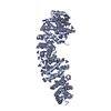
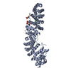
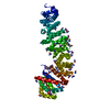
| Title | Crystal structure of mouse catenin beta-59 in 2.4M urea. | ||||||
 Components Components | Catenin beta-1 | ||||||
 Keywords Keywords | CELL ADHESION / mouse catenin | ||||||
| Function / homology |  Function and homology information Function and homology informationlung cell differentiation / epicardium-derived cardiac vascular smooth muscle cell differentiation / mesenchyme morphogenesis / RUNX3 regulates WNT signaling / Regulation of CDH11 function / cardiac vascular smooth muscle cell differentiation / Regulation of MITF-M-dependent genes involved in cell cycle and proliferation / Beta-catenin phosphorylation cascade / Apoptotic cleavage of cell adhesion proteins / Disassembly of the destruction complex and recruitment of AXIN to the membrane ...lung cell differentiation / epicardium-derived cardiac vascular smooth muscle cell differentiation / mesenchyme morphogenesis / RUNX3 regulates WNT signaling / Regulation of CDH11 function / cardiac vascular smooth muscle cell differentiation / Regulation of MITF-M-dependent genes involved in cell cycle and proliferation / Beta-catenin phosphorylation cascade / Apoptotic cleavage of cell adhesion proteins / Disassembly of the destruction complex and recruitment of AXIN to the membrane / hair cycle process / TCF dependent signaling in response to WNT / LRR FLII-interacting protein 1 (LRRFIP1) activates type I IFN production / endoderm formation / mesenchyme development / trachea morphogenesis / Formation of the beta-catenin:TCF transactivating complex / positive regulation of epithelial cell differentiation / positive regulation of heparan sulfate proteoglycan biosynthetic process / lung induction / positive regulation of branching involved in lung morphogenesis / cranial ganglion development / renal vesicle formation / renal inner medulla development / renal outer medulla development / nephron tubule formation / beta-catenin-ICAT complex / genitalia morphogenesis / embryonic skeletal limb joint morphogenesis / canonical Wnt signaling pathway involved in mesenchymal stem cell differentiation / neural plate development / metanephros morphogenesis / Deactivation of the beta-catenin transactivating complex / glial cell fate determination / astrocyte-dopaminergic neuron signaling / oviduct development / beta-catenin-TCF7L2 complex / regulation of nephron tubule epithelial cell differentiation / regulation of timing of anagen / negative regulation of mitotic cell cycle, embryonic / VEGFR2 mediated vascular permeability / regulation of secondary heart field cardioblast proliferation / negative regulation of mesenchymal to epithelial transition involved in metanephros morphogenesis / central nervous system vasculogenesis / regulation of centriole-centriole cohesion / regulation of epithelial cell differentiation / Adherens junctions interactions / regulation of centromeric sister chromatid cohesion / Degradation of beta-catenin by the destruction complex / embryonic axis specification / animal organ development / Ca2+ pathway / RHO GTPases activate IQGAPs / lens morphogenesis in camera-type eye / morphogenesis of embryonic epithelium / Scrib-APC-beta-catenin complex / beta-catenin-TCF complex / acinar cell differentiation / synaptic vesicle clustering / dorsal root ganglion development / endodermal cell fate commitment / neuron fate determination / proximal/distal pattern formation / endothelial tube morphogenesis / ventricular compact myocardium morphogenesis / dorsal/ventral axis specification / positive regulation of fibroblast growth factor receptor signaling pathway / sympathetic ganglion development / presynaptic active zone cytoplasmic component / fungiform papilla formation / positive regulation of endothelial cell differentiation / layer formation in cerebral cortex / mesenchymal to epithelial transition / hindbrain development / positive regulation of skeletal muscle tissue development / positive regulation of determination of dorsal identity / fascia adherens / lung epithelial cell differentiation / regulation of protein localization to cell surface / ectoderm development / embryonic foregut morphogenesis / cellular response to indole-3-methanol / positive regulation of odontoblast differentiation / mesenchymal cell proliferation involved in lung development / smooth muscle cell differentiation / positive regulation of myoblast proliferation / mesenchymal cell proliferation / alpha-catenin binding / histone methyltransferase binding / regulation of epithelial to mesenchymal transition / regulation of calcium ion import / cell projection membrane / regulation of osteoclast differentiation / positive regulation of homotypic cell-cell adhesion / negative regulation of oligodendrocyte differentiation / establishment of blood-retinal barrier / apicolateral plasma membrane / flotillin complex / epithelial cell differentiation involved in prostate gland development / positive regulation of epithelial cell proliferation involved in prostate gland development Similarity search - Function | ||||||
| Biological species |  | ||||||
| Method |  X-RAY DIFFRACTION / SYNCHROTRON / MOLECULAR REPLACEMENT / Resolution: 1.9 Å X-RAY DIFFRACTION / SYNCHROTRON / MOLECULAR REPLACEMENT / Resolution: 1.9 Å | ||||||
 Authors Authors | Wang, C. / Zhang, G.Y. | ||||||
 Citation Citation | Journal: TO BE PUBLISHED Title: Crystal structure of mouse catenin beta-59 in 2.4M urea. Authors: Wang, C. / Zhang, G.Y. | ||||||
| History |
|
- Structure visualization
| Structure viewer | Molecule: MolmilJmol/JSmol |
|---|
- Downloads & links
-Download
| PDBx/mmCIF format | 4ev8.cif.gz | 121.2 KB | Display | PDBx/mmCIF format |
|---|---|---|---|---|
| PDB format | pdb4ev8.ent.gz | 92.2 KB | Display | PDB format |
| PDBx/mmJSON format | 4ev8.json.gz | Tree view | PDBx/mmJSON format | |
| Others |  Other downloads Other downloads |
-Validation report
| Arichive directory | https://data.pdbj.org/pub/pdb/validation_reports/ev/4ev8ftp://data.pdbj.org/pub/pdb/validation_reports/ev/4ev8 | HTTPS FTP |
|---|
-Related structure data
| Related structure data |  3bctS S: Starting model for refinement |
|---|---|
| Similar structure data |


-Links
 PDBj
PDBj


- Assembly
| Deposited unit | 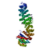
| ||||||||||||||||||
|---|---|---|---|---|---|---|---|---|---|---|---|---|---|---|---|---|---|---|---|
| 1 |
| ||||||||||||||||||
| Unit cell |
| ||||||||||||||||||
| Components on special symmetry positions |
|
-Components
| #1: Protein | Mass: 58844.117 Da / Num. of mol.: 1 Source method: isolated from a genetically manipulated source Source: (gene. exp.)  | ||
|---|---|---|---|
| #2: Chemical | ChemComp-URE /   Mass: 60.055 Da / Num. of mol.: 18 / Source method: obtained synthetically / Formula: CH4N2O Mass: 60.055 Da / Num. of mol.: 18 / Source method: obtained synthetically / Formula: CH4N2O#3: Water | ChemComp-HOH / |  Mass: 18.015 Da / Num. of mol.: 512 / Source method: isolated from a natural source / Formula: H2O Mass: 18.015 Da / Num. of mol.: 512 / Source method: isolated from a natural source / Formula: H2O |
-Experimental details
-Experiment
| Experiment | Method: X-RAY DIFFRACTION / Number of used crystals: 1 |
|---|
- Sample preparation
| Crystal | Density Matthews: 2.62 Å3/Da / Density % sol: 53 % |
|---|
-Data collection
| Diffraction | Mean temperature: 100 K |
|---|---|
| Diffraction source | Source: SYNCHROTRON / Site: ALS  / Beamline: 8.2.2 / Wavelength: 1 / Beamline: 8.2.2 / Wavelength: 1 |
| Detector | Type: ADSC QUANTUM 315 / Detector: CCD / Date: Mar 10, 2010 |
| Radiation | Monochromator: 0.99 / Protocol: SINGLE WAVELENGTH / Monochromatic (M) / Laue (L): M / Scattering type: x-ray |
| Radiation wavelength | Wavelength: 1 Å / Relative weight: 1 |
| Reflection | Resolution: 1.9→35.5 Å / Num. obs: 48834 / % possible obs: 96.4 % / Observed criterion σ(I): 1 / Redundancy: 6.2 % / Rsym value: 0.072 / Net I/σ(I): 26.1 |
| Reflection shell | Resolution: 1.9→1.94 Å / Rmerge(I) obs: 0.68 / Mean I/σ(I) obs: 2.7 / % possible all: 90.3 |
- Processing
| Software |
| ||||||||||||||||||||||||||||||||||||||||||||||||||||||||||||||||||||||||||||||||||||||||||||||||||||||||||||||||||||||||||||||
|---|---|---|---|---|---|---|---|---|---|---|---|---|---|---|---|---|---|---|---|---|---|---|---|---|---|---|---|---|---|---|---|---|---|---|---|---|---|---|---|---|---|---|---|---|---|---|---|---|---|---|---|---|---|---|---|---|---|---|---|---|---|---|---|---|---|---|---|---|---|---|---|---|---|---|---|---|---|---|---|---|---|---|---|---|---|---|---|---|---|---|---|---|---|---|---|---|---|---|---|---|---|---|---|---|---|---|---|---|---|---|---|---|---|---|---|---|---|---|---|---|---|---|---|---|---|---|---|
| Refinement | Method to determine structure: MOLECULAR REPLACEMENT Starting model: PDB ENTRY 3BCT Resolution: 1.9→35.47 Å / SU ML: 0.22 / σ(F): 1.34 / Phase error: 21.23 / Stereochemistry target values: ML
| ||||||||||||||||||||||||||||||||||||||||||||||||||||||||||||||||||||||||||||||||||||||||||||||||||||||||||||||||||||||||||||||
| Solvent computation | Shrinkage radii: 0.86 Å / VDW probe radii: 1.1 Å / Solvent model: FLAT BULK SOLVENT MODEL / Bsol: 60.82 Å2 / ksol: 0.34 e/Å3 | ||||||||||||||||||||||||||||||||||||||||||||||||||||||||||||||||||||||||||||||||||||||||||||||||||||||||||||||||||||||||||||||
| Displacement parameters |
| ||||||||||||||||||||||||||||||||||||||||||||||||||||||||||||||||||||||||||||||||||||||||||||||||||||||||||||||||||||||||||||||
| Refinement step | Cycle: LAST / Resolution: 1.9→35.47 Å
| ||||||||||||||||||||||||||||||||||||||||||||||||||||||||||||||||||||||||||||||||||||||||||||||||||||||||||||||||||||||||||||||
| Refine LS restraints |
| ||||||||||||||||||||||||||||||||||||||||||||||||||||||||||||||||||||||||||||||||||||||||||||||||||||||||||||||||||||||||||||||
| LS refinement shell |
|
