 Movie
Movie Controller

[English] 日本語
 Yorodumi
- PDB-4e4v: The crystal structure of the dimeric human importin alpha 1 at 2.... -
+ Open data

- Basic information
| Entry | Database: PDB / ID: 4e4v | ||||||
|---|---|---|---|---|---|---|---|
| Title | The crystal structure of the dimeric human importin alpha 1 at 2.5 angstrom resolution. | ||||||
 Components Components | Importin subunit alpha-2 | ||||||
 Keywords Keywords | TRANSPORT PROTEIN / armadillo repeat / karyopherin / importin / nuclear import / host-virus interaction / nucleus / phosphoprotein / protein transport / transport | ||||||
| Function / homology |  Function and homology information Function and homology informationSensing of DNA Double Strand Breaks / regulation of DNA recombination / entry of viral genome into host nucleus through nuclear pore complex via importin / positive regulation of viral life cycle / NS1 Mediated Effects on Host Pathways / NLS-dependent protein nuclear import complex / host cell / nuclear import signal receptor activity / CREB1 phosphorylation through the activation of CaMKII/CaMKK/CaMKIV cascasde / nuclear localization sequence binding ...Sensing of DNA Double Strand Breaks / regulation of DNA recombination / entry of viral genome into host nucleus through nuclear pore complex via importin / positive regulation of viral life cycle / NS1 Mediated Effects on Host Pathways / NLS-dependent protein nuclear import complex / host cell / nuclear import signal receptor activity / CREB1 phosphorylation through the activation of CaMKII/CaMKK/CaMKIV cascasde / nuclear localization sequence binding / DNA metabolic process / CaMK IV-mediated phosphorylation of CREB / NLS-bearing protein import into nucleus / ISG15 antiviral mechanism / protein import into nucleus / histone deacetylase binding / SARS-CoV-1 activates/modulates innate immune responses / nuclear membrane / Estrogen-dependent gene expression / Golgi membrane / endoplasmic reticulum membrane / SARS-CoV-2 activates/modulates innate and adaptive immune responses / RNA binding / nucleoplasm / membrane / nucleus / cytosol / cytoplasmSimilarity search - Function | ||||||
| Biological species |  Homo sapiens (human) Homo sapiens (human) | ||||||
| Method | X-RAY DIFFRACTION / SYNCHROTRON / MOLECULAR REPLACEMENT / Resolution: 2.5283 Å | ||||||
 Authors Authors | Hang, P.C. / Miknis, Z.M. / Franke, W.A. / Umland, T.C. / Schultz, L.W. | ||||||
 Citation Citation | Journal: To be Published Title: The crystal structure of the dimeric human importin alpha 1 at 2.5 angstrom resolution. Authors: Hang, P.C. / Miknis, Z.J. / Franke, W.A. / Umland, T.C. / Schultz, L.W. | ||||||
| History |
|
- Structure visualization
| Structure viewer | Molecule: MolmilJmol/JSmol |
|---|
- Downloads & links
-Download
| PDBx/mmCIF format | 4e4v.cif.gz | 494.1 KB | Display | PDBx/mmCIF format |
|---|---|---|---|---|
| PDB format | pdb4e4v.ent.gz | 417.3 KB | Display | PDB format |
| PDBx/mmJSON format | 4e4v.json.gz | Tree view | PDBx/mmJSON format | |
| Others |  Other downloads Other downloads |
-Validation report
| Arichive directory | https://data.pdbj.org/pub/pdb/validation_reports/e4/4e4vftp://data.pdbj.org/pub/pdb/validation_reports/e4/4e4v | HTTPS FTP |
|---|
-Related structure data
| Related structure data |  1ejlS S: Starting model for refinement |
|---|---|
| Similar structure data |


-Links
 PDBj
PDBj


- Assembly
| Deposited unit | 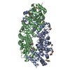
| ||||||||
|---|---|---|---|---|---|---|---|---|---|
| 1 | 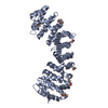
| ||||||||
| 2 | 
| ||||||||
| Unit cell |
|
-Components
| #1: Protein | / Karyopherin subunit alpha-2 / RAG cohort protein 1 / SRP1-alpha Mass: 52665.535 Da / Num. of mol.: 2 Fragment: Importin alpha 1 lacking IBB domain (UNP residues 70-529) Mutation: K486R Source method: isolated from a genetically manipulated source Source: (gene. exp.) Homo sapiens (human) / Gene: IP0A1, KPNA2, RCH1, SRP1 / Plasmid: PET15B / Production host:  Escherichia coli (E. coli) / Strain (production host): BL21 / References: UniProt: P52292 Escherichia coli (E. coli) / Strain (production host): BL21 / References: UniProt: P52292#2: Chemical | ChemComp-GOL / Glycerol  Mass: 92.094 Da / Num. of mol.: 13 / Source method: obtained synthetically / Formula: C3H8O3 Mass: 92.094 Da / Num. of mol.: 13 / Source method: obtained synthetically / Formula: C3H8O3#3: Chemical | Dithiothreitol  Mass: 154.251 Da / Num. of mol.: 2 / Source method: obtained synthetically / Formula: C4H10O2S2 Mass: 154.251 Da / Num. of mol.: 2 / Source method: obtained synthetically / Formula: C4H10O2S2#4: Water | ChemComp-HOH / | Water Mass: 18.015 Da / Num. of mol.: 166 / Source method: isolated from a natural source / Formula: H2O Mass: 18.015 Da / Num. of mol.: 166 / Source method: isolated from a natural source / Formula: H2O |
|---|
-Experimental details
-Experiment
| Experiment | Method: X-RAY DIFFRACTION / Number of used crystals: 1 |
|---|
- Sample preparation
| Crystal | Density Matthews: 3.35 Å3/Da / Density % sol: 63.26 % |
|---|---|
| Crystal grow | Temperature: 298 K / Method: vapor diffusion, sitting drop / pH: 5.4 Details: 0.5 CITRATE PH 5.4 10 MM DTT, VAPOR DIFFUSION, SITTING DROP, temperature 298K |
-Data collection
| Diffraction | Mean temperature: 100 K |
|---|---|
| Diffraction source | Source: SYNCHROTRON / Site: SSRL  / Beamline: BL9-1 / Wavelength: 0.9422 / Wavelength: 0.9422 Å / Beamline: BL9-1 / Wavelength: 0.9422 / Wavelength: 0.9422 Å |
| Detector | Type: ADSC QUANTUM 315r / Detector: CCD / Date: Jun 14, 2008 / Details: VERTICAL FOCUSING MIRROR |
| Radiation | Monochromator: SINGLE CRYSTAL SI(311) BENT MONOCHROMATOR (HORIZONTAL FOCUSING) Protocol: SINGLE WAVELENGTH / Monochromatic (M) / Laue (L): M / Scattering type: x-ray |
| Radiation wavelength | Wavelength: 0.9422 Å / Relative weight: 1 |
| Reflection | Resolution: 2.5→50 Å / Num. obs: 47596 / % possible obs: 99.7 % / Redundancy: 9.6 % / Biso Wilson estimate: 68.8 Å2 / Rmerge(I) obs: 0.057 / Net I/σ(I): 22.9 |
| Reflection shell | Resolution: 2.5→2.59 Å / Redundancy: 8.1 % / Rmerge(I) obs: 0.378 / Mean I/σ(I) obs: 5.02 / % possible all: 97.8 |
- Processing
| Software |
| |||||||||||||||||||||||||||||||||||||||||||||||||||||||||||||||||||||||||||||||||||||||||||||||||||||||||||||||||||||||||||||
|---|---|---|---|---|---|---|---|---|---|---|---|---|---|---|---|---|---|---|---|---|---|---|---|---|---|---|---|---|---|---|---|---|---|---|---|---|---|---|---|---|---|---|---|---|---|---|---|---|---|---|---|---|---|---|---|---|---|---|---|---|---|---|---|---|---|---|---|---|---|---|---|---|---|---|---|---|---|---|---|---|---|---|---|---|---|---|---|---|---|---|---|---|---|---|---|---|---|---|---|---|---|---|---|---|---|---|---|---|---|---|---|---|---|---|---|---|---|---|---|---|---|---|---|---|---|---|
| Refinement | Method to determine structure: MOLECULAR REPLACEMENT Starting model: PDB ENTRY 1EJL Resolution: 2.5283→34.809 Å / SU ML: 0.28 / σ(F): 1.34 / Phase error: 22.75 / Stereochemistry target values: ML
| |||||||||||||||||||||||||||||||||||||||||||||||||||||||||||||||||||||||||||||||||||||||||||||||||||||||||||||||||||||||||||||
| Solvent computation | Shrinkage radii: 0.9 Å / VDW probe radii: 1.11 Å / Solvent model: FLAT BULK SOLVENT MODEL | |||||||||||||||||||||||||||||||||||||||||||||||||||||||||||||||||||||||||||||||||||||||||||||||||||||||||||||||||||||||||||||
| Displacement parameters | Biso mean: 74.44 Å2
| |||||||||||||||||||||||||||||||||||||||||||||||||||||||||||||||||||||||||||||||||||||||||||||||||||||||||||||||||||||||||||||
| Refinement step | Cycle: LAST / Resolution: 2.5283→34.809 Å
| |||||||||||||||||||||||||||||||||||||||||||||||||||||||||||||||||||||||||||||||||||||||||||||||||||||||||||||||||||||||||||||
| Refine LS restraints |
| |||||||||||||||||||||||||||||||||||||||||||||||||||||||||||||||||||||||||||||||||||||||||||||||||||||||||||||||||||||||||||||
| LS refinement shell |
| |||||||||||||||||||||||||||||||||||||||||||||||||||||||||||||||||||||||||||||||||||||||||||||||||||||||||||||||||||||||||||||
| Refinement TLS params. | Method: refined / Refine-ID: X-RAY DIFFRACTION
| |||||||||||||||||||||||||||||||||||||||||||||||||||||||||||||||||||||||||||||||||||||||||||||||||||||||||||||||||||||||||||||
| Refinement TLS group |
|
