 Movie
Movie Controller

[English] 日本語
 Yorodumi
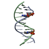
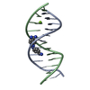
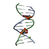
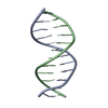
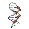
- PDB-4bna: REVERSIBLE BENDING AND HELIX GEOMETRY IN A B-DNA DODECAMER: CGCGA... -
+ Open data

- Basic information
| Entry | Database: PDB / ID: 4bna | ||||||||||||||||||
|---|---|---|---|---|---|---|---|---|---|---|---|---|---|---|---|---|---|---|---|
| Title | REVERSIBLE BENDING AND HELIX GEOMETRY IN A B-DNA DODECAMER: CGCGAATTBRCGCG | ||||||||||||||||||
 Components Components | DNA (5'-D(* Keywords KeywordsDNA / B-DNA / DOUBLE HELIX / MODIFIED | Function / homology | DNA / DNA (> 10) |  Function and homology information Function and homology informationMethod |  X-RAY DIFFRACTION / Resolution: 2.3 Å X-RAY DIFFRACTION / Resolution: 2.3 Å  Authors AuthorsKopka, M.L. / Fratini, A.V. / Dickerson, R.E. |  CitationJournal: J.Biol.Chem. / Year: 1982 CitationJournal: J.Biol.Chem. / Year: 1982Title: Reversible bending and helix geometry in a B-DNA dodecamer: CGCGAATTBrCGCG. Authors: Fratini, A.V. / Kopka, M.L. / Drew, H.R. / Dickerson, R.E. #1: Journal: J.Mol.Biol. / Year: 1983Title: Ordered Water Structure around a B-DNA Dodecamer. A Quantitative Study Authors: Kopka, M.L. / Fratini, A.V. / Drew, H.R. / Dickerson, R.E. #2: Journal: Proc.Natl.Acad.Sci.USA / Year: 1982Title: Structure of a B-DNA Dodecamer at 16 Kelvin Authors: Drew, H.R. / Samson, S. / Dickerson, R.E. #3: Journal: Proc.Natl.Acad.Sci.USA / Year: 1981Title: Kinematic Model for B-DNA Authors: Dickerson, R.E. / Drew, H.R. #4: Journal: Proc.Natl.Acad.Sci.USA / Year: 1981Title: Structure of a B-DNA Dodecamer. Conformation and Dynamics Authors: Drew, H.R. / Wing, R.M. / Takano, T. / Broka, C. / Tanaka, S. / Itakura, K. / Dickerson, R.E. #5: Journal: J.Mol.Biol. / Year: 1981Title: Structure of a B-DNA Dodecamer. II. Influence of Base Sequence on Helix Structure Authors: Dickerson, R.E. / Drew, H.R. #6: Journal: J.Mol.Biol. / Year: 1981Title: Structure of a B-DNA Dodecamer. III. Geometry of Hydration Authors: Drew, H.R. / Dickerson, R.E. #7: Journal: Nature / Year: 1980Title: Crystal Structure Analysis of a Complete Turn of B-DNA Authors: Wing, R. / Drew, H.R. / Takano, T. / Broka, C. / Tanaka, S. / Itakura, K. / Dickerson, R.E. History |
|
- Structure visualization
| Structure viewer | Molecule: MolmilJmol/JSmol |
|---|
- Downloads & links
-Download
| PDBx/mmCIF format | 4bna.cif.gz | 26.2 KB | Display | PDBx/mmCIF format |
|---|---|---|---|---|
| PDB format | pdb4bna.ent.gz | 17.3 KB | Display | PDB format |
| PDBx/mmJSON format | 4bna.json.gz | Tree view | PDBx/mmJSON format | |
| Others |  Other downloads Other downloads |
-Validation report
| Arichive directory | https://data.pdbj.org/pub/pdb/validation_reports/bn/4bnaftp://data.pdbj.org/pub/pdb/validation_reports/bn/4bna | HTTPS FTP |
|---|
-Related structure data


-Links
 PDBj
PDBj


- Assembly
| Deposited unit | 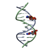
| ||||||||
|---|---|---|---|---|---|---|---|---|---|
| 1 |
| ||||||||
| Unit cell |
|
-Components
| #1: DNA chain | Mass: 3742.288 Da / Num. of mol.: 2 / Source method: obtained synthetically #2: Water | ChemComp-HOH / |  Mass: 18.015 Da / Num. of mol.: 114 / Source method: isolated from a natural source / Formula: H2O Mass: 18.015 Da / Num. of mol.: 114 / Source method: isolated from a natural source / Formula: H2O |
|---|
-Experimental details
-Experiment
| Experiment | Method: X-RAY DIFFRACTION |
|---|
- Sample preparation
| Crystal | Density Matthews: 2.07 Å3/Da / Density % sol: 40.65 % | |||||||||||||||||||||||||
|---|---|---|---|---|---|---|---|---|---|---|---|---|---|---|---|---|---|---|---|---|---|---|---|---|---|---|
| Crystal grow | Temperature: 280 K / Method: vapor diffusion / Details: VAPOR DIFFUSION, temperature 280.00K | |||||||||||||||||||||||||
| Components of the solutions |
| |||||||||||||||||||||||||
| Crystal grow | *PLUS Temperature: 7 ℃ | |||||||||||||||||||||||||
| Components of the solutions | *PLUS
|
-Data collection
| Diffraction | Mean temperature: 280 K |
|---|---|
| Detector | Detector: DIFFRACTOMETER |
| Radiation | Scattering type: x-ray |
| Radiation wavelength | Relative weight: 1 |
| Reflection | Resolution: 2.3→8 Å / Num. obs: 2919 |
| Reflection | *PLUS Highest resolution: 2.3 Å / Lowest resolution: 8 Å / Rmerge(I) obs: 0.59 |
- Processing
| Software | Name: JACK-LEVITT / Classification: refinement | ||||||||||||
|---|---|---|---|---|---|---|---|---|---|---|---|---|---|
| Refinement | Resolution: 2.3→8 Å /
| ||||||||||||
| Refine Biso |
| ||||||||||||
| Refinement step | Cycle: LAST / Resolution: 2.3→8 Å
| ||||||||||||
| Refinement | *PLUS Highest resolution: 2.3 Å / Lowest resolution: 7 Å / Rfactor obs: 0.215 | ||||||||||||
| Solvent computation | *PLUS | ||||||||||||
| Displacement parameters | *PLUS |
