- PDB-3zoo: Structure of the Y46F mutant of human cytochrome c -
+
Open data
ID or keywords:
Loading...
-
Basic information
Entry
Database: PDB / ID: 3zoo
Title
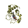
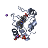
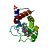
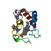
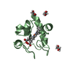
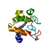
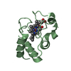
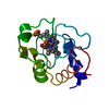
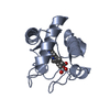
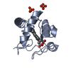
Structure of the Y46F mutant of human cytochrome c
Components
CYTOCHROME C
Keywords
OXIDOREDUCTASE / RESPIRATION / APOPTOSIS / ELECTRON TRANSFER
Function / homology
Function and homology information
Formation of apoptosome / apoptosome / Release of apoptotic factors from the mitochondria / Respiratory electron transport / cellular respiration / Activation of caspases through apoptosome-mediated cleavage / SMAC (DIABLO) binds to IAPs / SMAC(DIABLO)-mediated dissociation of IAP:caspase complexes / Regulation of the apoptosome activity / mitochondrial electron transport, cytochrome c to oxygen ...Formation of apoptosome / apoptosome / Release of apoptotic factors from the mitochondria / Respiratory electron transport / cellular respiration / Activation of caspases through apoptosome-mediated cleavage / SMAC (DIABLO) binds to IAPs / SMAC(DIABLO)-mediated dissociation of IAP:caspase complexes / Regulation of the apoptosome activity / mitochondrial electron transport, cytochrome c to oxygen / execution phase of apoptosis / mitochondrial electron transport, ubiquinol to cytochrome c / Detoxification of Reactive Oxygen Species / Pyroptosis / intrinsic apoptotic signaling pathway / TP53 Regulates Metabolic Genes / apoptotic signaling pathway / Transcriptional activation of mitochondrial biogenesis / Cytoprotection by HMOX1 / mitochondrial intermembrane space / electron transfer activity / mitochondrial inner membrane / heme binding / mitochondrion / metal ion binding / nucleus / cytosol Similarity search - Function
Resolution: 1.35→30.59 Å / Cor.coef. Fo:Fc: 0.978 / Cor.coef. Fo:Fc free: 0.968 / SU B: 2.408 / SU ML: 0.043 / Cross valid method: THROUGHOUT / ESU R: 0.056 / ESU R Free: 0.055 / Stereochemistry target values: MAXIMUM LIKELIHOOD / Details: HYDROGENS HAVE BEEN ADDED IN THE RIDING POSITIONS.
Rfactor
Num. reflection
% reflection
Selection details
Rfree
0.17935
4338
5 %
RANDOM
Rwork
0.13821
-
-
-
obs
0.14035
81707
94.73 %
-
Solvent computation
Ion probe radii: 0.8 Å / Shrinkage radii: 0.8 Å / VDW probe radii: 1.2 Å / Solvent model: MASK
 Movie
Movie Controller


 Open data
 Basic information
 Components
 Keywords
 Function and homology information
 HOMO SAPIENS (human)
 X-RAY DIFFRACTION /
  Authors
 Citation
 Structure visualization
 Downloads & links
 Other downloads


 PDBj
PDBj


 Assembly


 Mass: 618.503 Da / Num. of mol.: 4 / Source method: obtained synthetically / Formula: C34H34FeN4O4
 Mass: 94.971 Da / Num. of mol.: 2 / Source method: obtained synthetically / Formula: PO4
 Mass: 18.015 Da / Num. of mol.: 417 / Source method: isolated from a natural source / Formula: H2O
 Sample preparation
 / Beamline: I03 / Wavelength: 0.9762
  Processing