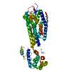
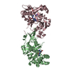
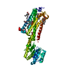
Entry Database : PDB / ID : 3vw7Title Crystal structure of human protease-activated receptor 1 (PAR1) bound with antagonist vorapaxar at 2.2 angstrom Proteinase-activated receptor 1, Lysozyme Keywords / / / / / / / / / Function / homology Function Domain/homology Component
/ / / / / / / / / / / / / / / / / / / / / / / / / / / / / / / / / / / / / / / / / / / / / / / / / / / / / / / / / / / / / / / / / / / / / / / / / / / / / / / / / / / / / / / / / / / / / / / / / / / / / / / / / Biological species Homo sapiens (human)Method / / / Resolution : 2.2 Å Authors Zhang, C. / Srinivasan, Y. / Arlow, D.H. / Fung, J.J. / Palmer, D. / Zheng, Y. / Green, H.F. / Pandey, A. / Dror, R.O. / Shaw, D.E. ...Zhang, C. / Srinivasan, Y. / Arlow, D.H. / Fung, J.J. / Palmer, D. / Zheng, Y. / Green, H.F. / Pandey, A. / Dror, R.O. / Shaw, D.E. / Weis, W.I. / Coughlin, S.R. / Kobilka, B.K. Journal : Nature / Year : 2012Title : High-resolution crystal structure of human protease-activated receptor 1Authors : Zhang, C. / Srinivasan, Y. / Arlow, D.H. / Fung, J.J. / Palmer, D. / Zheng, Y. / Green, H.F. / Pandey, A. / Dror, R.O. / Shaw, D.E. / Weis, W.I. / Coughlin, S.R. / Kobilka, B.K. History Deposition Aug 7, 2012 Deposition site / Processing site Revision 1.0 Dec 12, 2012 Provider / Type Revision 1.1 Aug 14, 2013 Group Revision 1.2 Aug 16, 2017 Group / Source and taxonomy / Category / pdbx_unobs_or_zero_occ_atomsRevision 1.3 Nov 22, 2017 Group / Category Item _software.classification / _software.contact_author ... _software.classification / _software.contact_author / _software.contact_author_email / _software.date / _software.language / _software.location / _software.name / _software.type / _software.version Revision 1.4 Nov 8, 2023 Group Advisory / Data collection ... Advisory / Data collection / Database references / Derived calculations / Refinement description / Structure summary Category chem_comp / chem_comp_atom ... chem_comp / chem_comp_atom / chem_comp_bond / database_2 / entity / pdbx_entity_nonpoly / pdbx_initial_refinement_model / pdbx_struct_conn_angle / pdbx_unobs_or_zero_occ_atoms / struct_conn / struct_ref_seq_dif / struct_site Item _chem_comp.name / _database_2.pdbx_DOI ... _chem_comp.name / _database_2.pdbx_DOI / _database_2.pdbx_database_accession / _entity.pdbx_description / _pdbx_entity_nonpoly.name / _pdbx_struct_conn_angle.ptnr1_auth_comp_id / _pdbx_struct_conn_angle.ptnr1_auth_seq_id / _pdbx_struct_conn_angle.ptnr1_label_asym_id / _pdbx_struct_conn_angle.ptnr1_label_atom_id / _pdbx_struct_conn_angle.ptnr1_label_comp_id / _pdbx_struct_conn_angle.ptnr1_label_seq_id / _pdbx_struct_conn_angle.ptnr3_auth_comp_id / _pdbx_struct_conn_angle.ptnr3_auth_seq_id / _pdbx_struct_conn_angle.ptnr3_label_asym_id / _pdbx_struct_conn_angle.ptnr3_label_atom_id / _pdbx_struct_conn_angle.ptnr3_label_comp_id / _pdbx_struct_conn_angle.ptnr3_label_seq_id / _pdbx_struct_conn_angle.value / _struct_conn.pdbx_dist_value / _struct_conn.ptnr1_auth_comp_id / _struct_conn.ptnr1_auth_seq_id / _struct_conn.ptnr1_label_asym_id / _struct_conn.ptnr1_label_atom_id / _struct_conn.ptnr1_label_comp_id / _struct_conn.ptnr1_label_seq_id / _struct_conn.ptnr2_auth_comp_id / _struct_conn.ptnr2_auth_seq_id / _struct_conn.ptnr2_label_asym_id / _struct_conn.ptnr2_label_atom_id / _struct_conn.ptnr2_label_comp_id / _struct_ref_seq_dif.details / _struct_site.pdbx_auth_asym_id / _struct_site.pdbx_auth_comp_id / _struct_site.pdbx_auth_seq_id Revision 1.5 Nov 20, 2024 Group / Category / pdbx_modification_feature
Show all Show less
 Movie
Movie Controller

 Yorodumi
 Open data
 Basic information
 Components
 Keywords
 Function and homology information
 Homo sapiens (human)
 Enterobacteria phage T4 (virus)
 X-RAY DIFFRACTION /
  Authors
 Citation
 Structure visualization
 Downloads & links
 Other downloads
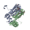

 PDBj
PDBj


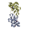
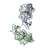
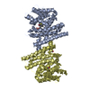
 Assembly

 Spodoptera frugiperda (fall armyworm) / References: UniProt: P25116, UniProt: P00720, lysozyme
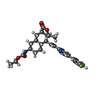


 Mass: 492.582 Da / Num. of mol.: 1 / Source method: obtained synthetically / Formula: C29H33FN2O4 / Comment: antagonist*YM
 Mass: 356.540 Da / Num. of mol.: 9 / Source method: obtained synthetically / Formula: C21H40O4
 Mass: 35.453 Da / Num. of mol.: 1 / Source method: obtained synthetically / Formula: Cl
 Mass: 22.990 Da / Num. of mol.: 1 / Source method: obtained synthetically / Formula: Na
 Sample preparation
 / Beamline: 23-ID-B / Wavelength: 1.033 Å
 Processing