 Movie
Movie Controller

[English] 日本語
 Yorodumi
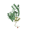
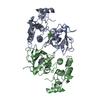
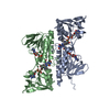
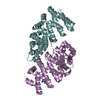
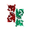
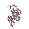
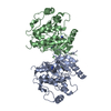
- PDB-3vht: Crystal structure of GFP-Wrnip1 UBZ domain fusion protein in comp... -
+ Open data

- Basic information
| Entry | Database: PDB / ID: 3vht | ||||||
|---|---|---|---|---|---|---|---|
| Title | Crystal structure of GFP-Wrnip1 UBZ domain fusion protein in complex with ubiquitin | ||||||
 Components Components |
| ||||||
 Keywords Keywords | FLUORESCENT PROTEIN/PROTEIN BINDING / GREEN FLUORESCENT PROTEIN / FUSION PROTEIN / ZINC FINGER / UBIQUITIN-BINDING DOMAIN / FLUORESCENT PROTEIN-PROTEIN BINDING complex | ||||||
| Function / homology |  Function and homology information Function and homology informationFormation of the ternary complex, and subsequently, the 43S complex / APC/C:Cdc20 mediated degradation of Cyclin B / SCF-beta-TrCP mediated degradation of Emi1 / APC-Cdc20 mediated degradation of Nek2A / ER Quality Control Compartment (ERQC) / Regulation of PTEN localization / Regulation of pyruvate metabolism / Downregulation of ERBB2:ERBB3 signaling / IRAK2 mediated activation of TAK1 complex / SMAD2/SMAD3:SMAD4 heterotrimer regulates transcription ...Formation of the ternary complex, and subsequently, the 43S complex / APC/C:Cdc20 mediated degradation of Cyclin B / SCF-beta-TrCP mediated degradation of Emi1 / APC-Cdc20 mediated degradation of Nek2A / ER Quality Control Compartment (ERQC) / Regulation of PTEN localization / Regulation of pyruvate metabolism / Downregulation of ERBB2:ERBB3 signaling / IRAK2 mediated activation of TAK1 complex / SMAD2/SMAD3:SMAD4 heterotrimer regulates transcription / PTK6 Regulates RTKs and Their Effectors AKT1 and DOK1 / Gap-filling DNA repair synthesis and ligation in GG-NER / Fanconi Anemia Pathway / Endosomal Sorting Complex Required For Transport (ESCRT) / Negative regulation of FLT3 / Synthesis of active ubiquitin: roles of E1 and E2 enzymes / Regulation of expression of SLITs and ROBOs / IRAK1 recruits IKK complex / IRAK1 recruits IKK complex upon TLR7/8 or 9 stimulation / Downregulation of ERBB4 signaling / E3 ubiquitin ligases ubiquitinate target proteins / Downregulation of TGF-beta receptor signaling / TGF-beta receptor signaling in EMT (epithelial to mesenchymal transition) / Stabilization of p53 / NOTCH3 Activation and Transmission of Signal to the Nucleus / Negative regulators of DDX58/IFIH1 signaling / Alpha-protein kinase 1 signaling pathway / Pexophagy / JNK (c-Jun kinases) phosphorylation and activation mediated by activated human TAK1 / Translesion synthesis by REV1 / Downregulation of SMAD2/3:SMAD4 transcriptional activity / Negative regulation of FGFR3 signaling / Negative regulation of FGFR4 signaling / Translesion synthesis by POLK / Formation of a pool of free 40S subunits / Regulation of NF-kappa B signaling / Negative regulation of FGFR1 signaling / Negative regulation of FGFR2 signaling / Regulation of TP53 Activity through Methylation / NRIF signals cell death from the nucleus / Translesion synthesis by POLI / Regulation of BACH1 activity / Recognition of DNA damage by PCNA-containing replication complex / SRP-dependent cotranslational protein targeting to membrane / p75NTR recruits signalling complexes / Interferon alpha/beta signaling / Negative regulation of MAPK pathway / Major pathway of rRNA processing in the nucleolus and cytosol / Spry regulation of FGF signaling / Regulation of TP53 Degradation / Nonsense Mediated Decay (NMD) independent of the Exon Junction Complex (EJC) / Translesion Synthesis by POLH / Activated NOTCH1 Transmits Signal to the Nucleus / Formation of TC-NER Pre-Incision Complex / Negative regulation of MET activity / TRAF6-mediated induction of TAK1 complex within TLR4 complex / IRAK2 mediated activation of TAK1 complex upon TLR7/8 or 9 stimulation / Termination of translesion DNA synthesis / Autodegradation of Cdh1 by Cdh1:APC/C / APC/C:Cdc20 mediated degradation of Securin / Senescence-Associated Secretory Phenotype (SASP) / Josephin domain DUBs / DNA Damage Recognition in GG-NER / Dual Incision in GG-NER / Ubiquitin-Mediated Degradation of Phosphorylated Cdc25A / Ubiquitin-dependent degradation of Cyclin D / Regulation of TBK1, IKKε (IKBKE)-mediated activation of IRF3, IRF7 / AUF1 (hnRNP D0) binds and destabilizes mRNA / Downregulation of ERBB2 signaling / Nonsense Mediated Decay (NMD) enhanced by the Exon Junction Complex (EJC) / Dual incision in TC-NER / Oncogene Induced Senescence / PINK1-PRKN Mediated Mitophagy / Cdc20:Phospho-APC/C mediated degradation of Cyclin A / SCF(Skp2)-mediated degradation of p27/p21 / N-glycan trimming in the ER and Calnexin/Calreticulin cycle / TNFR1-induced NF-kappa-B signaling pathway / Assembly of the pre-replicative complex / CDK-mediated phosphorylation and removal of Cdc6 / HDR through Homologous Recombination (HRR) / Gap-filling DNA repair synthesis and ligation in TC-NER / Translation initiation complex formation / Ribosomal scanning and start codon recognition / Inactivation of CSF3 (G-CSF) signaling / TCF dependent signaling in response to WNT / Metalloprotease DUBs / Formation of Incision Complex in GG-NER / Activation of IRF3, IRF7 mediated by TBK1, IKKε (IKBKE) / EGFR downregulation / Autodegradation of the E3 ubiquitin ligase COP1 / MAP3K8 (TPL2)-dependent MAPK1/3 activation / G2/M Checkpoints / Degradation of AXIN / Regulation of FZD by ubiquitination / Regulation of TNFR1 signaling / Asymmetric localization of PCP proteins / APC/C:Cdh1 mediated degradation of Cdc20 and other APC/C:Cdh1 targeted proteins in late mitosis/early G1 / Regulation of RUNX3 expression and activity / Regulation of RAS by GAPs / Regulation of PTEN stability and activity Similarity search - Function | ||||||
| Biological species |   Aequorea victoria (jellyfish) Aequorea victoria (jellyfish) Homo sapiens (human) Homo sapiens (human) | ||||||
| Method |  X-RAY DIFFRACTION / SYNCHROTRON / MOLECULAR REPLACEMENT / Resolution: 2.4 Å X-RAY DIFFRACTION / SYNCHROTRON / MOLECULAR REPLACEMENT / Resolution: 2.4 Å | ||||||
 Authors Authors | Suzuki, N. / Wakatsuki, S. / Kawasaki, M. | ||||||
 Citation Citation | Journal: Febs J. / Year: 2016 Title: A novel mode of ubiquitin recognition by the ubiquitin-binding zinc finger domain of WRNIP1. Authors: Suzuki, N. / Rohaim, A. / Kato, R. / Dikic, I. / Wakatsuki, S. / Kawasaki, M. | ||||||
| History |
|
- Structure visualization
| Structure viewer | Molecule: MolmilJmol/JSmol |
|---|
- Downloads & links
-Download
| PDBx/mmCIF format | 3vht.cif.gz | 236.9 KB | Display | PDBx/mmCIF format |
|---|---|---|---|---|
| PDB format | pdb3vht.ent.gz | 192.4 KB | Display | PDB format |
| PDBx/mmJSON format | 3vht.json.gz | Tree view | PDBx/mmJSON format | |
| Others |  Other downloads Other downloads |
-Validation report
| Arichive directory | https://data.pdbj.org/pub/pdb/validation_reports/vh/3vhtftp://data.pdbj.org/pub/pdb/validation_reports/vh/3vht | HTTPS FTP |
|---|
-Related structure data
| Related structure data | 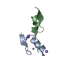 3vhsC  3wupC 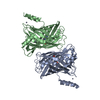 4z4kC 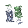 4z4mC 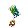 3ai5S C: citing same article ( S: Starting model for refinement |
|---|---|
| Similar structure data |

-Links
 PDBj
PDBj


- Assembly
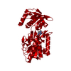
| Deposited unit | 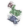
| ||||||||
|---|---|---|---|---|---|---|---|---|---|
| 1 |
| ||||||||
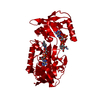
| Unit cell |
|
-Components
| #1: Protein | Mass: 26306.564 Da / Num. of mol.: 1 / Fragment: GFP domain Source method: isolated from a genetically manipulated source Source: (gene. exp.) Aequorea victoria (jellyfish) / Gene: GFP / Plasmid: pET28 / Production host:  |
|---|---|
| #2: Protein | Mass: 30543.525 Da / Num. of mol.: 1 / Fragment: GFP,Wrnip1 UBZ domain (UNP RESIDUES 9-46) Source method: isolated from a genetically manipulated source Details: THE FUSION PROTEIN OF YEAST ENHANCED GREEN FLUORESCENT PROTEIN, LINKER (GLY-SER) AND HUMAN WRNIP1 UBZ Source: (gene. exp.) Aequorea victoria (jellyfish), (gene. exp.) Homo sapiens (human)Gene: GFP, WRNIP1, WHIP / Plasmid: pET28 / Production host: References: UniProt: Q96S55, UniProt: P42212*PLUS, EC: 3.6.1.3 |
| #3: Protein | Mass: 8576.831 Da / Num. of mol.: 1 / Source method: isolated from a natural source / Source: (natural) |
| #4: Chemical | ChemComp-ZN /   Mass: 65.409 Da / Num. of mol.: 1 / Source method: obtained synthetically / Formula: Zn Mass: 65.409 Da / Num. of mol.: 1 / Source method: obtained synthetically / Formula: Zn |
| #5: Water | ChemComp-HOH /  Mass: 18.015 Da / Num. of mol.: 133 / Source method: isolated from a natural source / Formula: H2O Mass: 18.015 Da / Num. of mol.: 133 / Source method: isolated from a natural source / Formula: H2O |
| Has protein modification | Y |
| Sequence details | (1) THE SEQUENCE OF YEAST ENHANCED GREEN FLUORESCENT PROTEIN NOT CURRENTLY EXIST IN UNIPROT. THIS ...(1) THE SEQUENCE OF YEAST ENHANCED GREEN FLUORESCEN |
-Experimental details
-Experiment
| Experiment | Method: X-RAY DIFFRACTION / Number of used crystals: 1 |
|---|
- Sample preparation
| Crystal | Density Matthews: 2.61 Å3/Da / Density % sol: 52.86 % |
|---|---|
| Crystal grow | Temperature: 293 K / Method: vapor diffusion, sitting drop / pH: 7 Details: 0.2M DL-Malic acid, 20% PEG 3350, pH 7.0, VAPOR DIFFUSION, SITTING DROP, temperature 293.0K |
-Data collection
| Diffraction | Mean temperature: 100 K |
|---|---|
| Diffraction source | Source: SYNCHROTRON / Site: Photon Factory  / Beamline: AR-NW12A / Wavelength: 1 Å / Beamline: AR-NW12A / Wavelength: 1 Å |
| Detector | Type: ADSC QUANTUM 210 / Detector: CCD / Date: Dec 17, 2010 |
| Radiation | Monochromator: Si 111 / Protocol: SINGLE WAVELENGTH / Monochromatic (M) / Laue (L): M / Scattering type: x-ray |
| Radiation wavelength | Wavelength: 1 Å / Relative weight: 1 |
| Reflection | Resolution: 2.4→75.63 Å / Num. obs: 26943 / % possible obs: 99.4 % / Redundancy: 5.8 % / Biso Wilson estimate: 43.2 Å2 / Rmerge(I) obs: 0.105 / Net I/σ(I): 21.9 |
| Reflection shell | Resolution: 2.4→2.44 Å / Redundancy: 5.8 % / Rmerge(I) obs: 0.534 / Mean I/σ(I) obs: 3.7 / Num. unique all: 1328 / % possible all: 99.8 |
- Processing
| Software |
| ||||||||||||||||||||||||||||||||||||||||||||||||||||||||||||||||||||||||||||||||||||||||||||||||||||||||||||||||||||||||||||||||||||||||||||||||||||||||||||||||||||||||||
|---|---|---|---|---|---|---|---|---|---|---|---|---|---|---|---|---|---|---|---|---|---|---|---|---|---|---|---|---|---|---|---|---|---|---|---|---|---|---|---|---|---|---|---|---|---|---|---|---|---|---|---|---|---|---|---|---|---|---|---|---|---|---|---|---|---|---|---|---|---|---|---|---|---|---|---|---|---|---|---|---|---|---|---|---|---|---|---|---|---|---|---|---|---|---|---|---|---|---|---|---|---|---|---|---|---|---|---|---|---|---|---|---|---|---|---|---|---|---|---|---|---|---|---|---|---|---|---|---|---|---|---|---|---|---|---|---|---|---|---|---|---|---|---|---|---|---|---|---|---|---|---|---|---|---|---|---|---|---|---|---|---|---|---|---|---|---|---|---|---|---|---|
| Refinement | Method to determine structure: MOLECULAR REPLACEMENT Starting model: PDB ENTRY 3AI5 Resolution: 2.4→50 Å / Cor.coef. Fo:Fc: 0.946 / Cor.coef. Fo:Fc free: 0.914 / SU B: 16.892 / SU ML: 0.178 / Cross valid method: THROUGHOUT / ESU R Free: 0.248 / Stereochemistry target values: MAXIMUM LIKELIHOOD
| ||||||||||||||||||||||||||||||||||||||||||||||||||||||||||||||||||||||||||||||||||||||||||||||||||||||||||||||||||||||||||||||||||||||||||||||||||||||||||||||||||||||||||
| Solvent computation | Ion probe radii: 0.8 Å / Shrinkage radii: 0.8 Å / VDW probe radii: 1.4 Å / Solvent model: MASK | ||||||||||||||||||||||||||||||||||||||||||||||||||||||||||||||||||||||||||||||||||||||||||||||||||||||||||||||||||||||||||||||||||||||||||||||||||||||||||||||||||||||||||
| Displacement parameters | Biso mean: 32.581 Å2
| ||||||||||||||||||||||||||||||||||||||||||||||||||||||||||||||||||||||||||||||||||||||||||||||||||||||||||||||||||||||||||||||||||||||||||||||||||||||||||||||||||||||||||
| Refinement step | Cycle: LAST / Resolution: 2.4→50 Å
| ||||||||||||||||||||||||||||||||||||||||||||||||||||||||||||||||||||||||||||||||||||||||||||||||||||||||||||||||||||||||||||||||||||||||||||||||||||||||||||||||||||||||||
| Refine LS restraints |
| ||||||||||||||||||||||||||||||||||||||||||||||||||||||||||||||||||||||||||||||||||||||||||||||||||||||||||||||||||||||||||||||||||||||||||||||||||||||||||||||||||||||||||
| LS refinement shell | Resolution: 2.4→2.462 Å / Total num. of bins used: 20
| ||||||||||||||||||||||||||||||||||||||||||||||||||||||||||||||||||||||||||||||||||||||||||||||||||||||||||||||||||||||||||||||||||||||||||||||||||||||||||||||||||||||||||
| Refinement TLS params. | Method: refined / Origin x: -14.8285 Å / Origin y: -56.4302 Å / Origin z: -10.6721 Å
| ||||||||||||||||||||||||||||||||||||||||||||||||||||||||||||||||||||||||||||||||||||||||||||||||||||||||||||||||||||||||||||||||||||||||||||||||||||||||||||||||||||||||||
| Refinement TLS group |
|
