 Movie
Movie Controller

[English] 日本語
 Yorodumi
- PDB-3u9q: Ligand binding domain of PPARgamma complexed with Decanoic Acid a... -
+ Open data

- Basic information
| Entry | Database: PDB / ID: 3u9q | ||||||
|---|---|---|---|---|---|---|---|
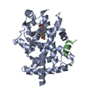
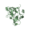
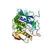
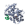
| Title | Ligand binding domain of PPARgamma complexed with Decanoic Acid and PGC-1a peptide | ||||||
 Components Components |
| ||||||
 Keywords Keywords |  TRANSCRIPTION / Nuclear receptor / Adipogenesis / RXRa / Nucleus TRANSCRIPTION / Nuclear receptor / Adipogenesis / RXRa / Nucleus | ||||||
| Function / homology |  Function and homology information Function and homology informationpositive regulation of mitochondrial DNA metabolic process / positive regulation of muscle tissue development / positive regulation of cellular respiration / positive regulation of fatty acid oxidation / : / : / lncRNA binding / cellular respiration / prostaglandin receptor activity / regulation of cholesterol transporter activity ...positive regulation of mitochondrial DNA metabolic process / positive regulation of muscle tissue development / positive regulation of cellular respiration / positive regulation of fatty acid oxidation / : / : / lncRNA binding / cellular respiration / prostaglandin receptor activity / regulation of cholesterol transporter activity / negative regulation of connective tissue replacement involved in inflammatory response wound healing / negative regulation of receptor signaling pathway via STAT / response to muscle activity / MECP2 regulates transcription factors / negative regulation of extracellular matrix assembly / negative regulation of vascular endothelial cell proliferation / negative regulation of cellular response to transforming growth factor beta stimulus / Activation of PPARGC1A (PGC-1alpha) by phosphorylation / negative regulation of cardiac muscle hypertrophy in response to stress / arachidonic acid binding / positive regulation of low-density lipoprotein receptor activity / positive regulation of adiponectin secretion / lipoprotein transport / negative regulation of sequestering of triglyceride / temperature homeostasis / positive regulation of vascular associated smooth muscle cell apoptotic process / macrophage derived foam cell differentiation / DNA binding domain binding / STAT family protein binding / positive regulation of fatty acid metabolic process / response to lipid / negative regulation of SMAD protein signal transduction / response to starvation / positive regulation of ATP biosynthetic process / LBD domain binding / negative regulation of type II interferon-mediated signaling pathway / negative regulation of cholesterol storage / response to dietary excess / E-box binding / alpha-actinin binding / negative regulation of blood vessel endothelial cell migration / lipid homeostasis / intracellular glucose homeostasis / fatty acid oxidation / negative regulation of vascular associated smooth muscle cell proliferation / R-SMAD binding / monocyte differentiation / positive regulation of cholesterol efflux / negative regulation of macrophage derived foam cell differentiation / cellular response to low-density lipoprotein particle stimulus / negative regulation of lipid storage / negative regulation of BMP signaling pathway / white fat cell differentiation / negative regulation of mitochondrial fission / positive regulation of fat cell differentiation / negative regulation of osteoblast differentiation / long-chain fatty acid transport / retinoic acid receptor signaling pathway / positive regulation of DNA binding / cell fate commitment / BMP signaling pathway / adipose tissue development / FOXO-mediated transcription of oxidative stress, metabolic and neuronal genes / nuclear retinoid X receptor binding / energy homeostasis / brown fat cell differentiation / negative regulation of signaling receptor activity / regulation of cellular response to insulin stimulus / cell maturation / positive regulation of gluconeogenesis / digestion / positive regulation of adipose tissue development / epithelial cell differentiation / respiratory electron transport chain / peroxisome proliferator activated receptor signaling pathway / hormone-mediated signaling pathway / mitochondrion organization / negative regulation of angiogenesis / response to nutrient / negative regulation of MAP kinase activity / RNA splicing / SUMOylation of transcription cofactors / fatty acid metabolic process / nuclear receptor coactivator activity / negative regulation of miRNA transcription / placenta development / Regulation of PTEN gene transcription / negative regulation of smooth muscle cell proliferation / gluconeogenesis / transcription coregulator binding / nuclear receptor binding / transcription initiation at RNA polymerase II promoter / transcription coregulator activity / peptide binding / negative regulation of transforming growth factor beta receptor signaling pathway / circadian regulation of gene expression / SUMOylation of intracellular receptors / Heme signaling / mRNA transcription by RNA polymerase II / Transcriptional activation of mitochondrial biogenesisSimilarity search - Function | ||||||
| Biological species |  Homo sapiens (human) Homo sapiens (human) | ||||||
| Method | X-RAY DIFFRACTION / SYNCHROTRON / MOLECULAR REPLACEMENT / Resolution: 1.522 Å | ||||||
 Authors Authors | Malapaka, V.R. / Xu, H.E. | ||||||
 Citation Citation | Journal: J.Biol.Chem. / Year: 2012 Title: Identification and Mechanism of 10-Carbon Fatty Acid as Modulating Ligand of Peroxisome Proliferator-activated Receptors. Authors: Malapaka, R.R. / Khoo, S. / Zhang, J. / Choi, J.H. / Zhou, X.E. / Xu, Y. / Gong, Y. / Li, J. / Yong, E.L. / Chalmers, M.J. / Chang, L. / Resau, J.H. / Griffin, P.R. / Chen, Y.E. / Xu, H.E. | ||||||
| History |
|
- Structure visualization
| Structure viewer | Molecule: MolmilJmol/JSmol |
|---|
- Downloads & links
-Download
| PDBx/mmCIF format | 3u9q.cif.gz | 121.4 KB | Display | PDBx/mmCIF format |
|---|---|---|---|---|
| PDB format | pdb3u9q.ent.gz | 93 KB | Display | PDB format |
| PDBx/mmJSON format | 3u9q.json.gz | Tree view | PDBx/mmJSON format | |
| Others |  Other downloads Other downloads |
-Validation report
| Arichive directory | https://data.pdbj.org/pub/pdb/validation_reports/u9/3u9qftp://data.pdbj.org/pub/pdb/validation_reports/u9/3u9q | HTTPS FTP |
|---|
-Related structure data
| Similar structure data |
|---|


-Links
 PDBj
PDBj


- Assembly
| Deposited unit | 
| ||||||||
|---|---|---|---|---|---|---|---|---|---|
| 1 |
| ||||||||
| Unit cell |
|
-Components
| #1: Protein | / PPAR-gamma / Nuclear receptor subfamily 1 group C member 3 Mass: 30704.734 Da / Num. of mol.: 1 / Fragment: Ligand Binding Domain, UNP residues 236-504 Source method: isolated from a genetically manipulated source Source: (gene. exp.) Homo sapiens (human) / Gene: NR1C3, PPARG / Production host:  Escherichia coli (E. coli) / Strain (production host): BL21(DE3) / References: UniProt: P37231 Escherichia coli (E. coli) / Strain (production host): BL21(DE3) / References: UniProt: P37231 |
|---|---|
| #2: Protein/peptide | Mass: 1000.319 Da / Num. of mol.: 1 / Fragment: Peptide, UNP residues 142-150 / Source method: obtained synthetically / Details: Synthetic peptide / Source: (synth.) Homo sapiens (human) / References: UniProt: Q9UBK2 |
| #3: Chemical | ChemComp-DKA / Capric acid  Mass: 172.265 Da / Num. of mol.: 1 / Source method: obtained synthetically / Formula: C10H20O2 Mass: 172.265 Da / Num. of mol.: 1 / Source method: obtained synthetically / Formula: C10H20O2 |
| #4: Water | ChemComp-HOH / Water Mass: 18.015 Da / Num. of mol.: 394 / Source method: isolated from a natural source / Formula: H2O Mass: 18.015 Da / Num. of mol.: 394 / Source method: isolated from a natural source / Formula: H2O |
-Experimental details
-Experiment
| Experiment | Method: X-RAY DIFFRACTION / Number of used crystals: 1 |
|---|
- Sample preparation
| Crystal | Density Matthews: 2.5 Å3/Da / Density % sol: 50.82 % |
|---|---|
| Crystal grow | Temperature: 298 K / Method: vapor diffusion / pH: 6.2 Details: 0.2 M sodium acetate (pH 6.2), 20% PEG 3350, 15% glycerol and 1 mM concentration of the ligand., VAPOR DIFFUSION, temperature 298K |
-Data collection
| Diffraction | Mean temperature: 200 K | |||||||||
|---|---|---|---|---|---|---|---|---|---|---|
| Diffraction source | Source: SYNCHROTRON / Site: APS  / Beamline: 21-ID-D / Wavelength: 0.62 - 1.91 / Beamline: 21-ID-D / Wavelength: 0.62 - 1.91 | |||||||||
| Detector | Type: MARMOSAIC 300 mm CCD / Detector: CCD / Date: Jun 6, 2009 | |||||||||
| Radiation | Monochromator: Ni filter / Protocol: SINGLE WAVELENGTH / Monochromatic (M) / Laue (L): M / Scattering type: x-ray | |||||||||
| Radiation wavelength |
| |||||||||
| Reflection | Resolution: 1.52→33.082 Å / Num. all: 45773 / Num. obs: 43465 / % possible obs: 94.9 % / Observed criterion σ(F): 2 / Observed criterion σ(I): 2 | |||||||||
| Reflection shell | Resolution: 1.522→1.562 Å / % possible all: 97.64 |
- Processing
| Software |
| ||||||||||||||||||||||||||||||||||||||||||||||||||||||||||||||||||||||||||||||||||||||||||||||||||||
|---|---|---|---|---|---|---|---|---|---|---|---|---|---|---|---|---|---|---|---|---|---|---|---|---|---|---|---|---|---|---|---|---|---|---|---|---|---|---|---|---|---|---|---|---|---|---|---|---|---|---|---|---|---|---|---|---|---|---|---|---|---|---|---|---|---|---|---|---|---|---|---|---|---|---|---|---|---|---|---|---|---|---|---|---|---|---|---|---|---|---|---|---|---|---|---|---|---|---|---|---|---|
| Refinement | Method to determine structure: MOLECULAR REPLACEMENT / Resolution: 1.522→30.082 Å / Cor.coef. Fo:Fc: 0.955 / Cor.coef. Fo:Fc free: 0.935 / Occupancy max: 1 / Occupancy min: 1 / SU B: 2.505 / SU ML: 0.044 / Cross valid method: THROUGHOUT / σ(F): 2 / ESU R Free: 0.083 / Stereochemistry target values: MAXIMUM LIKELIHOOD Details: HYDROGENS HAVE BEEN ADDED IN THE RIDING POSITIONS U VALUES : RESIDUAL ONLY
| ||||||||||||||||||||||||||||||||||||||||||||||||||||||||||||||||||||||||||||||||||||||||||||||||||||
| Solvent computation | Ion probe radii: 0.8 Å / Shrinkage radii: 0.8 Å / VDW probe radii: 1.4 Å / Solvent model: MASK | ||||||||||||||||||||||||||||||||||||||||||||||||||||||||||||||||||||||||||||||||||||||||||||||||||||
| Displacement parameters | Biso max: 100 Å2 / Biso mean: 20.037 Å2 / Biso min: 2 Å2
| ||||||||||||||||||||||||||||||||||||||||||||||||||||||||||||||||||||||||||||||||||||||||||||||||||||
| Refinement step | Cycle: LAST / Resolution: 1.522→30.082 Å
| ||||||||||||||||||||||||||||||||||||||||||||||||||||||||||||||||||||||||||||||||||||||||||||||||||||
| Refine LS restraints |
| ||||||||||||||||||||||||||||||||||||||||||||||||||||||||||||||||||||||||||||||||||||||||||||||||||||
| LS refinement shell | Resolution: 1.522→1.562 Å / Total num. of bins used: 20
| ||||||||||||||||||||||||||||||||||||||||||||||||||||||||||||||||||||||||||||||||||||||||||||||||||||
| Refinement TLS params. | Method: refined / Refine-ID: X-RAY DIFFRACTION
| ||||||||||||||||||||||||||||||||||||||||||||||||||||||||||||||||||||||||||||||||||||||||||||||||||||
| Refinement TLS group |
|
