 Movie
Movie Controller

[English] 日本語
 Yorodumi
- PDB-3sc1: Novel Isoquinolone PDK1 Inhibitors Discovered through Fragment-Ba... -
+ Open data

- Basic information
| Entry | Database: PDB / ID: 3sc1 | ||||||
|---|---|---|---|---|---|---|---|
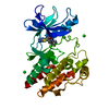
| Title | Novel Isoquinolone PDK1 Inhibitors Discovered through Fragment-Based Lead Discovery | ||||||
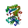
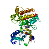
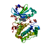
 Components Components | 3-phosphoinositide-dependent protein kinase 1 | ||||||
 Keywords Keywords | TRANSFERASE/TRANSFERASE INHIBITOR /  KINASE DOMAIN / PHOSPHOSERINE / SEP / TRANSFERASE-TRANSFERASE INHIBITOR complex KINASE DOMAIN / PHOSPHOSERINE / SEP / TRANSFERASE-TRANSFERASE INHIBITOR complex | ||||||
| Function / homology |  Function and homology information Function and homology information3-phosphoinositide-dependent protein kinase activity / Activation of AKT2 / regulation of mast cell degranulation / negative regulation of toll-like receptor signaling pathway / type B pancreatic cell development / positive regulation of phospholipase activity / RSK activation / hyperosmotic response / regulation of canonical NF-kappaB signal transduction / negative regulation of cardiac muscle cell apoptotic process ...3-phosphoinositide-dependent protein kinase activity / Activation of AKT2 / regulation of mast cell degranulation / negative regulation of toll-like receptor signaling pathway / type B pancreatic cell development / positive regulation of phospholipase activity / RSK activation / hyperosmotic response / regulation of canonical NF-kappaB signal transduction / negative regulation of cardiac muscle cell apoptotic process / positive regulation of vascular endothelial cell proliferation / phospholipase activator activity / positive regulation of sprouting angiogenesis / Constitutive Signaling by AKT1 E17K in Cancer / phospholipase binding / CD28 dependent PI3K/Akt signaling / positive regulation of blood vessel endothelial cell migration / Role of LAT2/NTAL/LAB on calcium mobilization / Estrogen-stimulated signaling through PRKCZ / SARS-CoV-2 targets host intracellular signalling and regulatory pathways / negative regulation of endothelial cell apoptotic process / SARS-CoV-1 targets host intracellular signalling and regulatory pathways / extrinsic apoptotic signaling pathway / RHO GTPases activate PKNs / cellular response to epidermal growth factor stimulus / GPVI-mediated activation cascade / T cell costimulation / activation of protein kinase B activity / Integrin signaling / positive regulation of release of sequestered calcium ion into cytosol / insulin-like growth factor receptor signaling pathway / VEGFR2 mediated vascular permeability / VEGFR2 mediated cell proliferation / cell projection / calcium-mediated signaling / positive regulation of protein localization to plasma membrane / negative regulation of transforming growth factor beta receptor signaling pathway / peptidyl-threonine phosphorylation / negative regulation of protein kinase activity / epidermal growth factor receptor signaling pathway / CLEC7A (Dectin-1) signaling / FCERI mediated NF-kB activation / G beta:gamma signalling through PI3Kgamma / cellular response to insulin stimulus / positive regulation of angiogenesis / cell migration / Regulation of TP53 Degradation / Downstream TCR signaling / PIP3 activates AKT signaling / insulin receptor signaling pathway / cytoplasmic vesicle / actin cytoskeleton organization / postsynaptic density / protein autophosphorylation / positive regulation of phosphatidylinositol 3-kinase/protein kinase B signal transduction / non-specific serine/threonine protein kinase / intracellular signal transduction / protein phosphorylation / focal adhesion / protein serine kinase activity / protein serine/threonine kinase activity / ATP binding / nucleus / plasma membrane / cytosol / cytoplasmSimilarity search - Function | ||||||
| Biological species |  Homo sapiens (human) Homo sapiens (human) | ||||||
| Method | X-RAY DIFFRACTION / SYNCHROTRON / FOURIER SYNTHESIS / Resolution: 2.7 Å | ||||||
 Authors Authors | Greasley, S.E. / Ferre, R.-A. / Krauss, M. / Cronin, C. | ||||||
 Citation Citation | Journal: J Comput Aided Mol Des / Year: 2011 Title: Novel isoquinolone PDK1 inhibitors discovered through fragment-based lead discovery. Authors: Johnson, M.C. / Hu, Q. / Lingardo, L. / Ferre, R.A. / Greasley, S. / Yan, J. / Kath, J. / Chen, P. / Ermolieff, J. / Alton, G. | ||||||
| History |
|
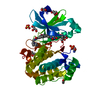
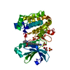
- Structure visualization
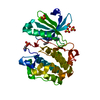
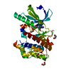
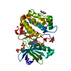
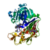
| Structure viewer | Molecule: MolmilJmol/JSmol |
|---|
- Downloads & links
-Download
| PDBx/mmCIF format | 3sc1.cif.gz | 67.3 KB | Display | PDBx/mmCIF format |
|---|---|---|---|---|
| PDB format | pdb3sc1.ent.gz | 53.6 KB | Display | PDB format |
| PDBx/mmJSON format | 3sc1.json.gz | Tree view | PDBx/mmJSON format | |
| Others |  Other downloads Other downloads |
-Validation report
| Arichive directory | https://data.pdbj.org/pub/pdb/validation_reports/sc/3sc1ftp://data.pdbj.org/pub/pdb/validation_reports/sc/3sc1 | HTTPS FTP |
|---|
-Related structure data
| Related structure data | |
|---|---|
| Similar structure data |


-Links
 PDBj
PDBj


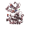
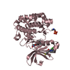
- Assembly
| Deposited unit | 
| ||||||||
|---|---|---|---|---|---|---|---|---|---|
| 1 |
| ||||||||
| 2 | 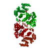
| ||||||||
| Unit cell |
|
-Components
| #1: Protein | Mass: 35555.738 Da / Num. of mol.: 1 / Fragment: Protein kinase domain, residues 50-359 Source method: isolated from a genetically manipulated source Source: (gene. exp.) Homo sapiens (human) / Gene: PDPK1, PDK1 / Production host:   Spodoptera frugiperda (fall armyworm) Spodoptera frugiperda (fall armyworm)References: UniProt: O15530, non-specific serine/threonine protein kinase | ||||
|---|---|---|---|---|---|
| #2: Chemical | ChemComp-3S1 / 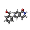  Mass: 251.280 Da / Num. of mol.: 1 / Source method: obtained synthetically / Formula: C16H13NO2 Mass: 251.280 Da / Num. of mol.: 1 / Source method: obtained synthetically / Formula: C16H13NO2 | ||||
| #3: Chemical | ChemComp-SO4 / Sulfate  Mass: 96.063 Da / Num. of mol.: 4 / Source method: obtained synthetically / Formula: SO4 Mass: 96.063 Da / Num. of mol.: 4 / Source method: obtained synthetically / Formula: SO4#4: Chemical | Glycerol  Mass: 92.094 Da / Num. of mol.: 2 / Source method: obtained synthetically / Formula: C3H8O3 Mass: 92.094 Da / Num. of mol.: 2 / Source method: obtained synthetically / Formula: C3H8O3#5: Water | ChemComp-HOH / | Water Mass: 18.015 Da / Num. of mol.: 31 / Source method: isolated from a natural source / Formula: H2O Mass: 18.015 Da / Num. of mol.: 31 / Source method: isolated from a natural source / Formula: H2O |
-Experimental details
-Experiment
| Experiment | Method: X-RAY DIFFRACTION / Number of used crystals: 1 |
|---|
- Sample preparation
| Crystal | Density Matthews: 2.94 Å3/Da / Density % sol: 58.14 % |
|---|---|
| Crystal grow | Temperature: 293 K / Method: vapor diffusion, hanging drop / pH: 7 Details: 2.2M AMMONIUM SULFATE, 10MM EDTA, 5-% GLYCEROL, 0.1M HEPES, pH 7.0, VAPOR DIFFUSION, HANGING DROP, temperature 293K |
-Data collection
| Diffraction | Mean temperature: 93 K |
|---|---|
| Diffraction source | Source: SYNCHROTRON / Site: ALS  / Beamline: 5.0.2 / Wavelength: 0.99 Å / Beamline: 5.0.2 / Wavelength: 0.99 Å |
| Detector | Type: ADSC QUANTUM 315 / Detector: CCD / Date: Jun 25, 2008 / Details: mirrors |
| Radiation | Monochromator: DOUBLE-CRYSTAL, SI(111) / Protocol: SINGLE WAVELENGTH / Monochromatic (M) / Laue (L): M / Scattering type: x-ray |
| Radiation wavelength | Wavelength: 0.99 Å / Relative weight: 1 |
| Reflection | Resolution: 2.65→50 Å / Num. all: 12487 / Num. obs: 12487 / % possible obs: 99.9 % / Observed criterion σ(F): 1 / Observed criterion σ(I): 1 / Redundancy: 4.13 % / Rsym value: 0.146 / Net I/σ(I): 11.9 |
| Reflection shell | Resolution: 2.65→2.7 Å / Redundancy: 3.4 % / Mean I/σ(I) obs: 1.3 / Num. unique all: 609 / Rsym value: 0.88 / % possible all: 99.8 |
- Processing
| Software |
| ||||||||||||||||||||
|---|---|---|---|---|---|---|---|---|---|---|---|---|---|---|---|---|---|---|---|---|---|
| Refinement | Method to determine structure: FOURIER SYNTHESIS / Resolution: 2.7→50 Å / σ(F): 0 / Stereochemistry target values: Engh & Huber
| ||||||||||||||||||||
| Refinement step | Cycle: LAST / Resolution: 2.7→50 Å
|
