 Movie
Movie Controller


+ Open data

- Basic information
| Entry | Database: PDB / ID: 3qtu | ||||||
|---|---|---|---|---|---|---|---|
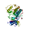
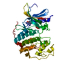
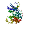
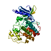
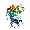
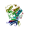
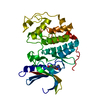
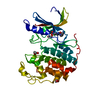
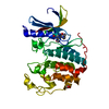
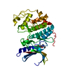
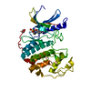
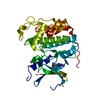
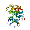
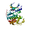
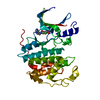
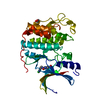
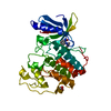
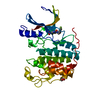
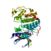
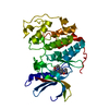
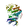
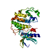
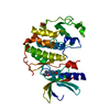
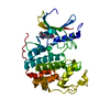
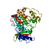
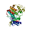
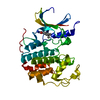
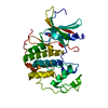
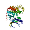
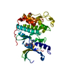
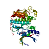
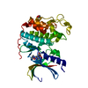
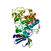
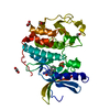
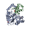
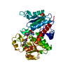
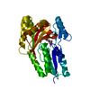
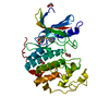
| Title | CDK2 in complex with inhibitor RC-2-132 | ||||||
 Components Components | Cyclin-dependent kinase 2 | ||||||
 Keywords Keywords | TRANSFERASE/TRANSFERASE INHIBITOR / protein kinase / inhibitor / TRANSFERASE-TRANSFERASE INHIBITOR complex | ||||||
| Function / homology |  Function and homology information Function and homology informationcyclin A1-CDK2 complex / cyclin E2-CDK2 complex / regulation of heterochromatin organization / cyclin E1-CDK2 complex / cyclin A2-CDK2 complex / positive regulation of DNA-templated DNA replication initiation / G2 Phase / Y chromosome / cyclin-dependent protein kinase activity / Phosphorylation of proteins involved in G1/S transition by active Cyclin E:Cdk2 complexes ...cyclin A1-CDK2 complex / cyclin E2-CDK2 complex / regulation of heterochromatin organization / cyclin E1-CDK2 complex / cyclin A2-CDK2 complex / positive regulation of DNA-templated DNA replication initiation / G2 Phase / Y chromosome / cyclin-dependent protein kinase activity / Phosphorylation of proteins involved in G1/S transition by active Cyclin E:Cdk2 complexes / positive regulation of heterochromatin formation / p53-Dependent G1 DNA Damage Response / X chromosome / PTK6 Regulates Cell Cycle / regulation of anaphase-promoting complex-dependent catabolic process / Defective binding of RB1 mutants to E2F1,(E2F2, E2F3) / centriole replication / Regulation of APC/C activators between G1/S and early anaphase / telomere maintenance in response to DNA damage / centrosome duplication / G0 and Early G1 / Telomere Extension By Telomerase / Activation of the pre-replicative complex / cyclin-dependent kinase / cyclin-dependent protein serine/threonine kinase activity / TP53 Regulates Transcription of Genes Involved in G1 Cell Cycle Arrest / Activation of ATR in response to replication stress / Regulation of MITF-M-dependent genes involved in cell cycle and proliferation / Cyclin E associated events during G1/S transition / Cajal body / Cyclin A:Cdk2-associated events at S phase entry / Cyclin A/B1/B2 associated events during G2/M transition / cyclin-dependent protein kinase holoenzyme complex / regulation of G2/M transition of mitotic cell cycle / condensed chromosome / negative regulation of protein localization to chromatin / mitotic G1 DNA damage checkpoint signaling / cellular response to nitric oxide / post-translational protein modification / regulation of mitotic cell cycle / cyclin binding / positive regulation of DNA replication / male germ cell nucleus / meiotic cell cycle / potassium ion transport / peptidyl-serine phosphorylation / G1/S transition of mitotic cell cycle / DNA Damage/Telomere Stress Induced Senescence / Meiotic recombination / CDK-mediated phosphorylation and removal of Cdc6 / G2/M transition of mitotic cell cycle / SCF(Skp2)-mediated degradation of p27/p21 / Transcriptional regulation of granulopoiesis / Orc1 removal from chromatin / Cyclin D associated events in G1 / cellular senescence / Regulation of TP53 Degradation / nuclear envelope / Factors involved in megakaryocyte development and platelet production / regulation of gene expression / Processing of DNA double-strand break ends / Senescence-Associated Secretory Phenotype (SASP) / transcription regulator complex / Regulation of TP53 Activity through Phosphorylation / Ras protein signal transduction / DNA replication / protein phosphorylation / chromosome, telomeric region / endosome / chromatin remodeling / protein domain specific binding / cell division / protein serine kinase activity / DNA repair / protein serine/threonine kinase activity / positive regulation of cell population proliferation / centrosome / DNA-templated transcription / positive regulation of DNA-templated transcription / magnesium ion binding / negative regulation of transcription by RNA polymerase II / signal transduction / nucleoplasm / ATP binding / nucleus / cytoplasm / cytosol Similarity search - Function | ||||||
| Biological species |  Homo sapiens (human) Homo sapiens (human) | ||||||
| Method |  X-RAY DIFFRACTION / MOLECULAR REPLACEMENT / Resolution: 1.82 Å X-RAY DIFFRACTION / MOLECULAR REPLACEMENT / Resolution: 1.82 Å | ||||||
 Authors Authors | Betzi, S. / Alam, R. / Han, H. / Becker, A. / Schonbrunn, E. | ||||||
 Citation Citation | Journal: J.Med.Chem. / Year: 2013 Title: Development of highly potent and selective diaminothiazole inhibitors of cyclin-dependent kinases. Authors: Schonbrunn, E. / Betzi, S. / Alam, R. / Martin, M.P. / Becker, A. / Han, H. / Francis, R. / Chakrasali, R. / Jakkaraj, S. / Kazi, A. / Sebti, S.M. / Cubitt, C.L. / Gebhard, A.W. / ...Authors: Schonbrunn, E. / Betzi, S. / Alam, R. / Martin, M.P. / Becker, A. / Han, H. / Francis, R. / Chakrasali, R. / Jakkaraj, S. / Kazi, A. / Sebti, S.M. / Cubitt, C.L. / Gebhard, A.W. / Hazlehurst, L.A. / Tash, J.S. / Georg, G.I. | ||||||
| History |
|
- Structure visualization
| Structure viewer | Molecule: MolmilJmol/JSmol |
|---|
- Downloads & links
-Download
| PDBx/mmCIF format | 3qtu.cif.gz | 78.4 KB | Display | PDBx/mmCIF format |
|---|---|---|---|---|
| PDB format | pdb3qtu.ent.gz | 57.6 KB | Display | PDB format |
| PDBx/mmJSON format | 3qtu.json.gz | Tree view | PDBx/mmJSON format | |
| Others |  Other downloads Other downloads |
-Validation report
| Arichive directory | https://data.pdbj.org/pub/pdb/validation_reports/qt/3qtuftp://data.pdbj.org/pub/pdb/validation_reports/qt/3qtu | HTTPS FTP |
|---|
-Related structure data
| Related structure data |  3qqkC 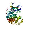 3qtqC 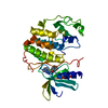 3qtrC 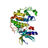 3qtsC 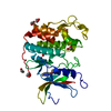 3qtwC 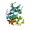 3qtxC 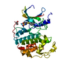 3qtzC  3qu0C  3qxpC  3r8uC 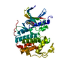 3r8vC 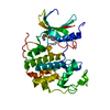 3r8zC 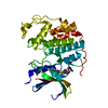 3r9dC 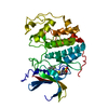 3r9hC  3r9nC 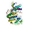 3r9oC 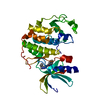 3rahC 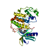 3rakC 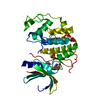 3ralC 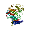 3rjcC 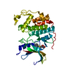 3rk5C 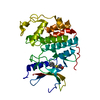 3rk7C 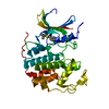 3rk9C 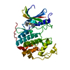 3rkbC 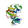 3rmfC 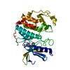 3rniC 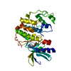 3rprC 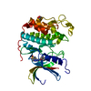 3rpvC  3rpyC 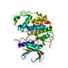 3rzbC 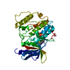 3s00C 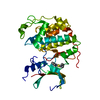 3s0oC 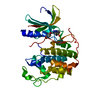 3s1hC 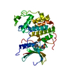 3sqqC 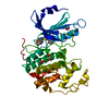 4gcjC 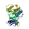 1pw2S C: citing same article ( S: Starting model for refinement |
|---|---|
| Similar structure data |


-Links
 PDBj
PDBj


- Assembly
| Deposited unit | 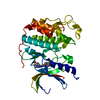
| ||||||||
|---|---|---|---|---|---|---|---|---|---|
| 1 |
| ||||||||
| Unit cell |
|
-Components
| #1: Protein | Mass: 34761.348 Da / Num. of mol.: 1 Source method: isolated from a genetically manipulated source Source: (gene. exp.) Homo sapiens (human) / Gene: CDK2 / Plasmid: pGEX6P-1 / Production host:  |
|---|---|
| #2: Chemical | ChemComp-X44 /   Mass: 453.516 Da / Num. of mol.: 1 / Source method: obtained synthetically / Formula: C16H15N5O5S3 Mass: 453.516 Da / Num. of mol.: 1 / Source method: obtained synthetically / Formula: C16H15N5O5S3 |
| #3: Water | ChemComp-HOH /  Mass: 18.015 Da / Num. of mol.: 133 / Source method: isolated from a natural source / Formula: H2O Mass: 18.015 Da / Num. of mol.: 133 / Source method: isolated from a natural source / Formula: H2O |
-Experimental details
-Experiment
| Experiment | Method: X-RAY DIFFRACTION / Number of used crystals: 1 |
|---|
- Sample preparation
| Crystal | Density Matthews: 2.03 Å3/Da / Density % sol: 39.34 % |
|---|---|
| Crystal grow | Temperature: 291 K / Method: vapor diffusion, hanging drop / pH: 7.5 Details: 5 mg/mL CDK2 protein, 3 mM inhibitor, 15% v/v PEG3350, 50 mM HEPES/NaOH, 50 mM Na/K phosphate, pH 7.5, VAPOR DIFFUSION, HANGING DROP, temperature 291K |
-Data collection
| Diffraction | Mean temperature: 93 K |
|---|---|
| Diffraction source | Source: ROTATING ANODE / Type: RIGAKU MICROMAX-007 HF / Wavelength: 1.54178 |
| Detector | Type: RIGAKU SATURN 944+ / Detector: CCD / Date: May 12, 2010 / Details: MIRRORS |
| Radiation | Monochromator: MIRRORS / Protocol: SINGLE WAVELENGTH / Monochromatic (M) / Laue (L): M / Scattering type: x-ray |
| Radiation wavelength | Wavelength: 1.54178 Å / Relative weight: 1 |
| Reflection | Resolution: 1.82→23.12 Å / Num. all: 25332 / Num. obs: 25332 / % possible obs: 97.4 % / Observed criterion σ(F): 0 / Observed criterion σ(I): -3 / Redundancy: 5.8 % / Biso Wilson estimate: 18.4 Å2 / Rmerge(I) obs: 0.053 / Net I/σ(I): 54.4 |
| Reflection shell | Resolution: 1.82→1.85 Å / Redundancy: 3.9 % / Rmerge(I) obs: 0.364 / Mean I/σ(I) obs: 5.95 / Num. unique all: 1186 / % possible all: 91.4 |
- Processing
| Software |
| ||||||||||||||||||||||||||||||||||||||||||||||||||||||||||||||||||||||||||||||||
|---|---|---|---|---|---|---|---|---|---|---|---|---|---|---|---|---|---|---|---|---|---|---|---|---|---|---|---|---|---|---|---|---|---|---|---|---|---|---|---|---|---|---|---|---|---|---|---|---|---|---|---|---|---|---|---|---|---|---|---|---|---|---|---|---|---|---|---|---|---|---|---|---|---|---|---|---|---|---|---|---|---|
| Refinement | Method to determine structure: MOLECULAR REPLACEMENT Starting model: PDB ENTRY 1PW2 Resolution: 1.82→23.12 Å / Rfactor Rfree error: 0.008 / Data cutoff high absF: 673394.25 / Data cutoff low absF: 0 / Isotropic thermal model: RESTRAINED / Cross valid method: THROUGHOUT / σ(F): 0 / Stereochemistry target values: Engh & Huber / Details: BULK SOLVENT MODEL USED
| ||||||||||||||||||||||||||||||||||||||||||||||||||||||||||||||||||||||||||||||||
| Solvent computation | Solvent model: FLAT MODEL / Bsol: 60.2802 Å2 / ksol: 0.4 e/Å3 | ||||||||||||||||||||||||||||||||||||||||||||||||||||||||||||||||||||||||||||||||
| Displacement parameters | Biso mean: 38.1 Å2
| ||||||||||||||||||||||||||||||||||||||||||||||||||||||||||||||||||||||||||||||||
| Refine analyze |
| ||||||||||||||||||||||||||||||||||||||||||||||||||||||||||||||||||||||||||||||||
| Refinement step | Cycle: LAST / Resolution: 1.82→23.12 Å
| ||||||||||||||||||||||||||||||||||||||||||||||||||||||||||||||||||||||||||||||||
| Refine LS restraints |
| ||||||||||||||||||||||||||||||||||||||||||||||||||||||||||||||||||||||||||||||||
| Refine LS restraints NCS | NCS model details: NONE | ||||||||||||||||||||||||||||||||||||||||||||||||||||||||||||||||||||||||||||||||
| LS refinement shell | Resolution: 1.82→1.93 Å / Rfactor Rfree error: 0.026 / Total num. of bins used: 6
| ||||||||||||||||||||||||||||||||||||||||||||||||||||||||||||||||||||||||||||||||
| Xplor file |
|
